 Movie
Movie Controller

[English] 日本語
 Yorodumi
- PDB-6b67: Human PP2Calpha (PPM1A) complexed with cyclic peptide c(MpSIpYVA) -
+ Open data

- Basic information
| Entry | Database: PDB / ID: 6b67 | ||||||
|---|---|---|---|---|---|---|---|
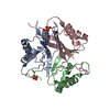
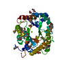
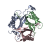
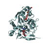
| Title | Human PP2Calpha (PPM1A) complexed with cyclic peptide c(MpSIpYVA) | ||||||
 Components Components |
| ||||||
 Keywords Keywords | HYDROLASE / Phosphatase | ||||||
| Function / homology |  Function and homology information Function and homology informationN-terminal protein myristoylation / calmodulin-dependent protein phosphatase activity / Energy dependent regulation of mTOR by LKB1-AMPK / negative regulation of non-canonical NF-kappaB signal transduction / regulation of canonical NF-kappaB signal transduction / protein dephosphorylation / protein-serine/threonine phosphatase / protein serine/threonine phosphatase activity / R-SMAD binding / negative regulation of BMP signaling pathway ...N-terminal protein myristoylation / calmodulin-dependent protein phosphatase activity / Energy dependent regulation of mTOR by LKB1-AMPK / negative regulation of non-canonical NF-kappaB signal transduction / regulation of canonical NF-kappaB signal transduction / protein dephosphorylation / protein-serine/threonine phosphatase / protein serine/threonine phosphatase activity / R-SMAD binding / negative regulation of BMP signaling pathway / cellular response to transforming growth factor beta stimulus / protein export from nucleus / positive regulation of protein export from nucleus / negative regulation of canonical NF-kappaB signal transduction / negative regulation of transforming growth factor beta receptor signaling pathway / Downregulation of SMAD2/3:SMAD4 transcriptional activity / positive regulation of canonical Wnt signaling pathway / manganese ion binding / positive regulation of canonical NF-kappaB signal transduction / regulation of cell cycle / positive regulation of DNA-templated transcription / magnesium ion binding / negative regulation of transcription by RNA polymerase II / nucleoplasm / membrane / nucleus / plasma membrane / cytosol Similarity search - Function | ||||||
| Biological species |  Homo sapiens (human) Homo sapiens (human)synthetic construct (others) | ||||||
| Method |  X-RAY DIFFRACTION / MOLECULAR REPLACEMENT / Resolution: 2.2 Å X-RAY DIFFRACTION / MOLECULAR REPLACEMENT / Resolution: 2.2 Å | ||||||
 Authors Authors | Dyda, F. / Kosek, D. | ||||||
 Citation Citation | Journal: J. Biol. Chem. / Year: 2018 Title: A trapped human PPM1A-phosphopeptide complex reveals structural features critical for regulation of PPM protein phosphatase activity. Authors: Debnath, S. / Kosek, D. / Tagad, H.D. / Durell, S.R. / Appella, D.H. / Acevedo, R. / Grishaev, A. / Dyda, F. / Appella, E. / Mazur, S.J. | ||||||
| History |
|
- Structure visualization
| Structure viewer | Molecule: MolmilJmol/JSmol |
|---|
- Downloads & links
-Download
| PDBx/mmCIF format | 6b67.cif.gz | 204.6 KB | Display | PDBx/mmCIF format |
|---|---|---|---|---|
| PDB format | pdb6b67.ent.gz | 162.1 KB | Display | PDB format |
| PDBx/mmJSON format | 6b67.json.gz | Tree view | PDBx/mmJSON format | |
| Others |  Other downloads Other downloads |
-Validation report
| Arichive directory | https://data.pdbj.org/pub/pdb/validation_reports/b6/6b67ftp://data.pdbj.org/pub/pdb/validation_reports/b6/6b67 | HTTPS FTP |
|---|
-Related structure data
| Related structure data | 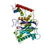 1a6qS S: Starting model for refinement |
|---|---|
| Similar structure data |


-Links
 PDBj
PDBj

- Assembly
| Deposited unit | 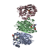
| ||||||||
|---|---|---|---|---|---|---|---|---|---|
| 1 | 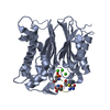
| ||||||||
| 2 | 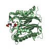
| ||||||||
| 3 | 
| ||||||||
| Unit cell |
|
-Components
| #1: Protein | Mass: 32845.914 Da / Num. of mol.: 3 / Mutation: D146E Source method: isolated from a genetically manipulated source Source: (gene. exp.) Homo sapiens (human) / Gene: PPM1A, PPPM1A / Production host:  References: UniProt: P35813, protein-serine/threonine phosphatase #2: Protein/peptide | Mass: 1002.981 Da / Num. of mol.: 3 / Source method: obtained synthetically Details: The cyclic peptide is a synthetic construct whose structure is represented in the attached files. We can also provide th is information in other formats, if this is more convenient. A ...Details: The cyclic peptide is a synthetic construct whose structure is represented in the attached files. We can also provide th is information in other formats, if this is more convenient. A general method for preparation of thioether cyclic peptid es is given by Long et al. (2003). The specific cyclic peptide, c(M-pS-I-pY-V-A) was first reported to be a substrate of PPM1A by Yamaguchi et al. 2006. Source: (synth.) synthetic construct (others) #3: Chemical | ChemComp-CA /   Mass: 40.078 Da / Num. of mol.: 10 / Source method: obtained synthetically / Formula: Ca Mass: 40.078 Da / Num. of mol.: 10 / Source method: obtained synthetically / Formula: Ca#4: Water | ChemComp-HOH / |  Mass: 18.015 Da / Num. of mol.: 633 / Source method: isolated from a natural source / Formula: H2O Mass: 18.015 Da / Num. of mol.: 633 / Source method: isolated from a natural source / Formula: H2O |
|---|
-Experimental details
-Experiment
| Experiment | Method: X-RAY DIFFRACTION / Number of used crystals: 1 |
|---|
- Sample preparation
| Crystal | Density Matthews: 2.26 Å3/Da / Density % sol: 45.55 % |
|---|---|
| Crystal grow | Temperature: 293 K / Method: vapor diffusion, hanging drop / pH: 7.3 Details: Protein conc: 20 mg/ml Protein buffer: 10 mM MES, pH 7.0, 150 mM NaCl, 2 mM TCEP and 2 mM MgCl2 Protein and cyclic peptide was dialyzed against above buffer and mixed at the 1:2 molar ratio ...Details: Protein conc: 20 mg/ml Protein buffer: 10 mM MES, pH 7.0, 150 mM NaCl, 2 mM TCEP and 2 mM MgCl2 Protein and cyclic peptide was dialyzed against above buffer and mixed at the 1:2 molar ratio Precipitant: 0.1M HEPES (pH 7.3), 0.1M calcium acetate and 40% PEG400 |
-Data collection
| Diffraction | Mean temperature: 95 K |
|---|---|
| Diffraction source | Source: ROTATING ANODE / Type: RIGAKU MICROMAX-007 HF / Wavelength: 1.5418 Å |
| Detector | Type: RIGAKU SATURN A200 / Detector: CCD / Date: Jul 6, 2016 |
| Radiation | Monochromator: Multilayer KB pair / Protocol: SINGLE WAVELENGTH / Monochromatic (M) / Laue (L): M / Scattering type: x-ray |
| Radiation wavelength | Wavelength: 1.5418 Å / Relative weight: 1 |
| Reflection | Resolution: 2.2→30 Å / Num. obs: 43867 / % possible obs: 95.7 % / Observed criterion σ(I): -3 / Redundancy: 2.87 % / Rsym value: 0.03 / Net I/σ(I): 25.09 |
| Reflection shell | Resolution: 2.2→2.26 Å / Mean I/σ(I) obs: 4.63 / Rsym value: 0.093 |
- Processing
| Software |
| ||||||||||||||||||||
|---|---|---|---|---|---|---|---|---|---|---|---|---|---|---|---|---|---|---|---|---|---|
| Refinement | Method to determine structure: MOLECULAR REPLACEMENT Starting model: 1A6Q Resolution: 2.2→30 Å / Cross valid method: FREE R-VALUE / σ(F): 0 Details: CARTESIAN SIMULATED ANNEALING, RESTRAINED APD REFINEMENT, ENERGY MIMIZATION
| ||||||||||||||||||||
| Refinement step | Cycle: LAST / Resolution: 2.2→30 Å
| ||||||||||||||||||||
| LS refinement shell | Resolution: 2.2→2.24 Å / Rfactor Rfree: 0.264 / Rfactor Rwork: 0.2177 |
