 Movie
Movie Controller

[English] 日本語
 Yorodumi
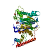
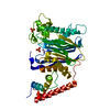
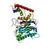
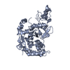
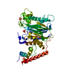
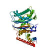
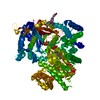
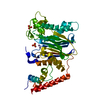
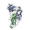
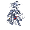
- PDB-1a6q: CRYSTAL STRUCTURE OF THE PROTEIN SERINE/THREONINE PHOSPHATASE 2C ... -
+ Open data

- Basic information
| Entry | Database: PDB / ID: 1a6q | |||||||||
|---|---|---|---|---|---|---|---|---|---|---|
| Title | CRYSTAL STRUCTURE OF THE PROTEIN SERINE/THREONINE PHOSPHATASE 2C AT 2 A RESOLUTION | |||||||||
 Components Components | PHOSPHATASE 2C | |||||||||
 Keywords Keywords | HYDROLASE / CATALYTIC MECHANISM / METALLOENZYME / PROTEIN PHOSPHATASE 2C / SIGNAL TRANSDUCTUIN | |||||||||
| Function / homology |  Function and homology information Function and homology informationN-terminal protein myristoylation / calmodulin-dependent protein phosphatase activity / Energy dependent regulation of mTOR by LKB1-AMPK / negative regulation of non-canonical NF-kappaB signal transduction / regulation of canonical NF-kappaB signal transduction / protein dephosphorylation / protein-serine/threonine phosphatase / protein serine/threonine phosphatase activity / R-SMAD binding / negative regulation of BMP signaling pathway ...N-terminal protein myristoylation / calmodulin-dependent protein phosphatase activity / Energy dependent regulation of mTOR by LKB1-AMPK / negative regulation of non-canonical NF-kappaB signal transduction / regulation of canonical NF-kappaB signal transduction / protein dephosphorylation / protein-serine/threonine phosphatase / protein serine/threonine phosphatase activity / R-SMAD binding / negative regulation of BMP signaling pathway / cellular response to transforming growth factor beta stimulus / protein export from nucleus / positive regulation of protein export from nucleus / negative regulation of canonical NF-kappaB signal transduction / negative regulation of transforming growth factor beta receptor signaling pathway / Downregulation of SMAD2/3:SMAD4 transcriptional activity / positive regulation of canonical Wnt signaling pathway / manganese ion binding / positive regulation of canonical NF-kappaB signal transduction / regulation of cell cycle / positive regulation of DNA-templated transcription / magnesium ion binding / negative regulation of transcription by RNA polymerase II / nucleoplasm / membrane / nucleus / plasma membrane / cytosol Similarity search - Function | |||||||||
| Biological species |  Homo sapiens (human) Homo sapiens (human) | |||||||||
| Method |  X-RAY DIFFRACTION / SYNCHROTRON / MIR / Resolution: 2 Å X-RAY DIFFRACTION / SYNCHROTRON / MIR / Resolution: 2 Å | |||||||||
 Authors Authors | Das, A.K. / Helps, N.R. / Cohen, P.T.W. / Barford, D. | |||||||||
 Citation Citation | Journal: EMBO J. / Year: 1996 Title: Crystal structure of the protein serine/threonine phosphatase 2C at 2.0 A resolution. Authors: Das, A.K. / Helps, N.R. / Cohen, P.T. / Barford, D. | |||||||||
| History |
|
- Structure visualization
| Structure viewer | Molecule: MolmilJmol/JSmol |
|---|
- Downloads & links
-Download
| PDBx/mmCIF format | 1a6q.cif.gz | 86.9 KB | Display | PDBx/mmCIF format |
|---|---|---|---|---|
| PDB format | pdb1a6q.ent.gz | 65.4 KB | Display | PDB format |
| PDBx/mmJSON format | 1a6q.json.gz | Tree view | PDBx/mmJSON format | |
| Others |  Other downloads Other downloads |
-Validation report
| Arichive directory | https://data.pdbj.org/pub/pdb/validation_reports/a6/1a6qftp://data.pdbj.org/pub/pdb/validation_reports/a6/1a6q | HTTPS FTP |
|---|
-Related structure data
| Similar structure data |
|---|
-Links
 PDBj
PDBj

- Assembly
| Deposited unit | 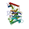
| ||||||||
|---|---|---|---|---|---|---|---|---|---|
| 1 |
| ||||||||
| Unit cell |
|
-Components
| #1: Protein | Mass: 42502.703 Da / Num. of mol.: 1 Source method: isolated from a genetically manipulated source Source: (gene. exp.) Homo sapiens (human) / Production host:  References: UniProt: P35813, protein-serine/threonine phosphatase | ||||
|---|---|---|---|---|---|
| #2: Chemical |   Mass: 54.938 Da / Num. of mol.: 2 / Source method: obtained synthetically / Formula: Mn Mass: 54.938 Da / Num. of mol.: 2 / Source method: obtained synthetically / Formula: Mn#3: Chemical | ChemComp-PO4 / |   Mass: 94.971 Da / Num. of mol.: 1 / Source method: obtained synthetically / Formula: PO4 Mass: 94.971 Da / Num. of mol.: 1 / Source method: obtained synthetically / Formula: PO4#4: Water | ChemComp-HOH / |  Mass: 18.015 Da / Num. of mol.: 203 / Source method: isolated from a natural source / Formula: H2O Mass: 18.015 Da / Num. of mol.: 203 / Source method: isolated from a natural source / Formula: H2O |
-Experimental details
-Experiment
| Experiment | Method: X-RAY DIFFRACTION / Number of used crystals: 5 |
|---|
- Sample preparation
| Crystal | Density Matthews: 2.97 Å3/Da / Density % sol: 59 % | ||||||||||||||||||||||||||||||||||||||||||||||||||||||||||||
|---|---|---|---|---|---|---|---|---|---|---|---|---|---|---|---|---|---|---|---|---|---|---|---|---|---|---|---|---|---|---|---|---|---|---|---|---|---|---|---|---|---|---|---|---|---|---|---|---|---|---|---|---|---|---|---|---|---|---|---|---|---|
| Crystal grow | Temperature: 277 K / pH: 5 Details: 8-12% PEG8K,15% GLYCEROL, 50MM K-PO4, PH=5.0, 2 MM DTT AT 4 DEGREES C, temperature 277K | ||||||||||||||||||||||||||||||||||||||||||||||||||||||||||||
| Crystal grow | *PLUS Temperature: 100 K / Method: vapor diffusionDetails: drop solution was mixed with an equal volume of reservoir solution | ||||||||||||||||||||||||||||||||||||||||||||||||||||||||||||
| Components of the solutions | *PLUS
|
-Data collection
| Diffraction | Mean temperature: 100 K |
|---|---|
| Diffraction source | Source: SYNCHROTRON / Site: ESRF  / Beamline: BL19 / Wavelength: 0.87 / Beamline: BL19 / Wavelength: 0.87 |
| Detector | Type: MARRESEARCH / Detector: IMAGE PLATE / Date: Feb 9, 1996 / Details: MIRRORS |
| Radiation | Monochromator: DARESBURY / Monochromatic (M) / Laue (L): M / Scattering type: x-ray |
| Radiation wavelength | Wavelength: 0.87 Å / Relative weight: 1 |
| Reflection | Resolution: 2→15 Å / Num. obs: 33176 / % possible obs: 95.5 % / Observed criterion σ(I): 2 / Redundancy: 6.5 % / Rmerge(I) obs: 0.089 / Rsym value: 0.089 / Net I/σ(I): 12.6 |
| Reflection | *PLUS Num. measured all: 215022 |
- Processing
| Software |
| ||||||||||||||||||||||||||||||||||||||||||||||||||||||||||||
|---|---|---|---|---|---|---|---|---|---|---|---|---|---|---|---|---|---|---|---|---|---|---|---|---|---|---|---|---|---|---|---|---|---|---|---|---|---|---|---|---|---|---|---|---|---|---|---|---|---|---|---|---|---|---|---|---|---|---|---|---|---|
| Refinement | Method to determine structure: MIR / Resolution: 2→6 Å / Data cutoff high absF: 10000000 / Data cutoff low absF: 0.001 / Cross valid method: THROUGHOUT / σ(F): 2
| ||||||||||||||||||||||||||||||||||||||||||||||||||||||||||||
| Refinement step | Cycle: LAST / Resolution: 2→6 Å
| ||||||||||||||||||||||||||||||||||||||||||||||||||||||||||||
| Refine LS restraints |
| ||||||||||||||||||||||||||||||||||||||||||||||||||||||||||||
| Xplor file |
| ||||||||||||||||||||||||||||||||||||||||||||||||||||||||||||
| Software | *PLUS Name: X-PLOR / Version: 3.82 / Classification: refinement | ||||||||||||||||||||||||||||||||||||||||||||||||||||||||||||
| Refinement | *PLUS | ||||||||||||||||||||||||||||||||||||||||||||||||||||||||||||
| Solvent computation | *PLUS | ||||||||||||||||||||||||||||||||||||||||||||||||||||||||||||
| Displacement parameters | *PLUS | ||||||||||||||||||||||||||||||||||||||||||||||||||||||||||||
| Refine LS restraints | *PLUS
|
