 Movie
Movie Controller


+ Open data

- Basic information
| Entry | Database: PDB / ID: 1hwu | ||||||
|---|---|---|---|---|---|---|---|
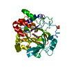
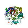
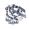
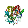
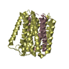
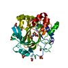
| Title | STRUCTURE OF PII PROTEIN FROM HERBASPIRILLUM SEROPEDICAE | ||||||
 Components Components | PII PROTEIN | ||||||
 Keywords Keywords | SIGNALING PROTEIN / Herbaspirillum seropedicae PII / Beta-alpha-beta motif / signal transduction protein | ||||||
| Function / homology |  Function and homology information Function and homology informationregulation of nitrogen utilization / enzyme regulator activity / DNA-templated transcription / ATP binding / cytosol Similarity search - Function | ||||||
| Biological species |  Herbaspirillum seropedicae (bacteria) Herbaspirillum seropedicae (bacteria) | ||||||
| Method |  X-RAY DIFFRACTION / SYNCHROTRON / MOLECULAR REPLACEMENT / Resolution: 2.1 Å X-RAY DIFFRACTION / SYNCHROTRON / MOLECULAR REPLACEMENT / Resolution: 2.1 Å | ||||||
 Authors Authors | Benelli, E.M. / Buck, M. / Polikarpov, I. / De Souza, E.M. / Cruz, L.M. / Pedrosa, F.O. | ||||||
 Citation Citation | Journal: Eur.J.Biochem. / Year: 2002 Title: Herbaspirillum seropedicae signal transduction protein PII is structurally similar to the enteric GlnK. Authors: Machado Benelli, E. / Buck, M. / Polikarpov, I. / Maltempi de Souza, E. / Cruz, L.M. / Pedrosa, F.O. | ||||||
| History |
|
- Structure visualization
| Structure viewer | Molecule: MolmilJmol/JSmol |
|---|
- Downloads & links
-Download
| PDBx/mmCIF format | 1hwu.cif.gz | 116.4 KB | Display | PDBx/mmCIF format |
|---|---|---|---|---|
| PDB format | pdb1hwu.ent.gz | 92.9 KB | Display | PDB format |
| PDBx/mmJSON format | 1hwu.json.gz | Tree view | PDBx/mmJSON format | |
| Others |  Other downloads Other downloads |
-Validation report
| Arichive directory | https://data.pdbj.org/pub/pdb/validation_reports/hw/1hwuftp://data.pdbj.org/pub/pdb/validation_reports/hw/1hwu | HTTPS FTP |
|---|
-Related structure data
| Related structure data |  2piiS S: Starting model for refinement |
|---|---|
| Similar structure data |


-Links
 PDBj
PDBj- Assembly
| Deposited unit | 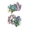
| ||||||||
|---|---|---|---|---|---|---|---|---|---|
| 1 | 
| ||||||||
| 2 | 
| ||||||||
| 3 | 
| ||||||||
| Unit cell |
|
-Components
| #1: Protein | Mass: 12284.189 Da / Num. of mol.: 6 Source method: isolated from a genetically manipulated source Source: (gene. exp.) Herbaspirillum seropedicae (bacteria) / Gene: GLNB / Plasmid: PET28B+ / Production host: #2: Water | ChemComp-HOH / |  Mass: 18.015 Da / Num. of mol.: 118 / Source method: isolated from a natural source / Formula: H2O Mass: 18.015 Da / Num. of mol.: 118 / Source method: isolated from a natural source / Formula: H2O |
|---|
-Experimental details
-Experiment
| Experiment | Method: X-RAY DIFFRACTION / Number of used crystals: 1 |
|---|
- Sample preparation
| Crystal | Density Matthews: 2.21 Å3/Da / Density % sol: 44.33 % | |||||||||||||||||||||||||||||||||||||||||||||||||||||||||||||||
|---|---|---|---|---|---|---|---|---|---|---|---|---|---|---|---|---|---|---|---|---|---|---|---|---|---|---|---|---|---|---|---|---|---|---|---|---|---|---|---|---|---|---|---|---|---|---|---|---|---|---|---|---|---|---|---|---|---|---|---|---|---|---|---|---|
| Crystal grow | Temperature: 291 K / Method: vapor diffusion, hanging drop / pH: 6.5 Details: Sodium Cacodylate, Magnesium Acetate, Methylpentadiol, pH 6.5, VAPOR DIFFUSION, HANGING DROP, temperature 291K | |||||||||||||||||||||||||||||||||||||||||||||||||||||||||||||||
| Crystal grow | *PLUS pH: 8 / Method: vapor diffusion, hanging drop | |||||||||||||||||||||||||||||||||||||||||||||||||||||||||||||||
| Components of the solutions | *PLUS
|
-Data collection
| Diffraction | Mean temperature: 120 K |
|---|---|
| Diffraction source | Source: SYNCHROTRON / Site: LNLS  / Beamline: D03B-MX1 / Wavelength: 1.38 Å / Beamline: D03B-MX1 / Wavelength: 1.38 Å |
| Detector | Type: MARRESEARCH / Detector: IMAGE PLATE / Date: Jun 18, 1998 |
| Radiation | Monochromator: Si 111 CHANNEL / Protocol: SINGLE WAVELENGTH / Monochromatic (M) / Laue (L): M / Scattering type: x-ray |
| Radiation wavelength | Wavelength: 1.38 Å / Relative weight: 1 |
| Reflection | Resolution: 2.1→13 Å / Num. all: 146413 / Num. obs: 36523 / % possible obs: 94.4 % / Observed criterion σ(I): 3 / Redundancy: 4 % / Rmerge(I) obs: 0.057 |
| Reflection shell | Resolution: 2.1→2.15 Å / Rmerge(I) obs: 0.322 / Num. unique all: 36523 / % possible all: 95.5 |
| Reflection | *PLUS % possible obs: 94 % / Redundancy: 3 % |
| Reflection shell | *PLUS Highest resolution: 2.1 Å / % possible obs: 95 % / Num. unique obs: 2415 |
- Processing
| Software |
| ||||||||||||||||||||
|---|---|---|---|---|---|---|---|---|---|---|---|---|---|---|---|---|---|---|---|---|---|
| Refinement | Method to determine structure: MOLECULAR REPLACEMENT Starting model: PDB ENTRY 2PII Resolution: 2.1→13 Å / Cross valid method: THROUGHOUT
| ||||||||||||||||||||
| Refinement step | Cycle: LAST / Resolution: 2.1→13 Å
| ||||||||||||||||||||
| Refine LS restraints |
| ||||||||||||||||||||
| Refinement | *PLUS Num. reflection obs: 36331 / Num. reflection Rfree: 1820 / % reflection Rfree: 5 % | ||||||||||||||||||||
| Solvent computation | *PLUS | ||||||||||||||||||||
| Displacement parameters | *PLUS |
