| Software | | Name | Version | Classification |
|---|
| HKL-2000 | | data scaling| PHENIX | 1.12_2829refinement| PDB_EXTRACT | 3.22 | data extraction| HKL-2000 | | data reduction| PHASER | | phasing | | | | | |
|
|---|
| Refinement | Resolution: 2.035→46.214 Å / Cross valid method: FREE R-VALUE / σ(F): 36.94 / Phase error: 43.08
| Rfactor | Num. reflection | % reflection |
|---|
| Rfree | 0.2696 | 2020 | 4.68 % |
|---|
| Rwork | 0.2502 | - | - |
|---|
| obs | 0.2601 | 43163 | 99.06 % |
|---|
|
|---|
| Solvent computation | Shrinkage radii: 0.9 Å / VDW probe radii: 1.11 Å |
|---|
| Displacement parameters | Biso max: 194.37 Å2 / Biso mean: 57.93 Å2 / Biso min: 29.74 Å2 |
|---|
| Refinement step | Cycle: final / Resolution: 2.035→46.214 Å
| Protein | Nucleic acid | Ligand | Solvent | Total |
|---|
| Num. atoms | 5542 | 0 | 0 | 76 | 5618 |
|---|
| Biso mean | - | - | - | 52.54 | - |
|---|
| Num. residues | - | - | - | - | 715 |
|---|
|
|---|
| Refine LS restraints | | Refine-ID | Type | Dev ideal | Number |
|---|
| X-RAY DIFFRACTION | f_bond_d| 0.004 | 5645 | | X-RAY DIFFRACTION | f_angle_d| 0.701 | 7666 | | X-RAY DIFFRACTION | f_chiral_restr| 0.093 | 868 | | X-RAY DIFFRACTION | f_plane_restr| 0.003 | 1014 | | X-RAY DIFFRACTION | f_dihedral_angle_d| 17.065 | 3384 | | | | | |
|
|---|
| LS refinement shell | Refine-ID: X-RAY DIFFRACTION / Rfactor Rfree error: 0 | Resolution (Å) | Rfactor Rfree | Num. reflection Rfree | Rfactor Rwork | Num. reflection Rwork | % reflection obs (%) |
|---|
| 2.0366-2.0876 | 0.3287 | 158 | 0.3418 | 2957 | 95 | | 2.0876-2.144 | 0.3729 | 124 | 0.3508 | 2989 | 96 | | 2.144-2.2071 | 0.4652 | 169 | 0.4214 | 2945 | 94 | | 2.2071-2.2783 | 0.4374 | 136 | 0.4334 | 2976 | 95 | | 2.2783-2.3597 | 0.4551 | 129 | 0.5072 | 2942 | 95 | | 2.3597-2.4541 | 0.3885 | 136 | 0.3607 | 2947 | 96 | | 2.4541-2.5657 | 0.3872 | 144 | 0.3774 | 2987 | 95 | | 2.5657-2.7009 | 0.3615 | 147 | 0.3468 | 2959 | 95 | | 2.7009-2.87 | 0.352 | 146 | 0.3213 | 2956 | 95 | | 2.87-3.0913 | 0.2782 | 130 | 0.2881 | 2963 | 96 | | 3.0913-3.402 | 0.2376 | 149 | 0.2478 | 2949 | 95 | | 3.402-3.8933 | 0.2251 | 147 | 0.2036 | 2957 | 95 | | 3.8933-4.9013 | 0.2034 | 160 | 0.1635 | 2884 | 93 | | 4.9013-29.1075 | 0.2921 | 138 | 0.2174 | 2684 | 86 |
|
|---|
| Refinement TLS params. | Method: refined / Origin x: -12.3948 Å / Origin y: 35.5099 Å / Origin z: 39.4326 Å
| 11 | 12 | 13 | 21 | 22 | 23 | 31 | 32 | 33 |
|---|
| T | 0.2949 Å2 | -0.0444 Å2 | 0.0118 Å2 | - | 0.3172 Å2 | 0.039 Å2 | - | - | 0.3783 Å2 |
|---|
| L | 0.6801 °2 | -0.0552 °2 | -0.0384 °2 | - | 0.7429 °2 | 0.0375 °2 | - | - | 0.5247 °2 |
|---|
| S | -0.008 Å ° | -0.0316 Å ° | -0.1566 Å ° | 0.0158 Å ° | -0.0209 Å ° | 0.0443 Å ° | 0.1625 Å ° | -0.1036 Å ° | 0.0437 Å ° |
|---|
|
|---|
| Refinement TLS group | | ID | Refine-ID | Refine TLS-ID | Selection details | Auth asym-ID | Auth seq-ID |
|---|
| 1 | X-RAY DIFFRACTION | 1 | allA| 29 - 454 | | 2 | X-RAY DIFFRACTION | 1 | allB| 30 - 454 | | 3 | X-RAY DIFFRACTION | 1 | allS| 1 - 76 | | | | | | |
|
|---|
 Movie
Movie Controller

 Yorodumi
 Open data
 Basic information
 Components
 Keywords
 Function and homology information
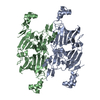
 Bertholletia excelsa (Brazil nut)
 X-RAY DIFFRACTION /
  Authors
 Citation
 Structure visualization
 Downloads & links
 Other downloads


 PDBj
PDBj Assembly
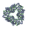
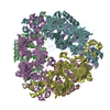
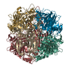

 Mass: 18.015 Da / Num. of mol.: 76 / Source method: isolated from a natural source / Formula: H2O
 Sample preparation
 Processing