 Movie
Movie Controller


+ Open data

- Basic information
| Entry | Database: PDB / ID: 5wpw | ||||||
|---|---|---|---|---|---|---|---|
| Title | Crystal structure of coconut allergen cocosin | ||||||
 Components Components | 11S globulin isoform 1 | ||||||
 Keywords Keywords | ALLERGEN / 11S globulin / seed storage protein | ||||||
| Function / homology |  Function and homology information Function and homology informationseed maturation / extraorganismal space / seed development / nutrient reservoir activity Similarity search - Function | ||||||
| Biological species |  Cocos nucifera (coconut palm) Cocos nucifera (coconut palm) | ||||||
| Method |  X-RAY DIFFRACTION / SYNCHROTRON / MOLECULAR REPLACEMENT / Resolution: 1.847 Å X-RAY DIFFRACTION / SYNCHROTRON / MOLECULAR REPLACEMENT / Resolution: 1.847 Å | ||||||
 Authors Authors | Jin, T. / Zhang, Y. | ||||||
 Citation Citation | Journal: J. Agric. Food Chem. / Year: 2017 Title: Crystal Structure of Cocosin, A Potential Food Allergen from Coconut (Cocos nucifera) Authors: Jin, T. / Wang, C. / Zhang, C. / Wang, Y. / Chen, Y.W. / Guo, F. / Howard, A. / Cao, M.J. / Fu, T.J. / McHugh, T.H. / Zhang, Y. | ||||||
| History |
|
- Structure visualization
| Structure viewer | Molecule: MolmilJmol/JSmol |
|---|
- Downloads & links
-Download
| PDBx/mmCIF format | 5wpw.cif.gz | 165.6 KB | Display | PDBx/mmCIF format |
|---|---|---|---|---|
| PDB format | pdb5wpw.ent.gz | 129.1 KB | Display | PDB format |
| PDBx/mmJSON format | 5wpw.json.gz | Tree view | PDBx/mmJSON format | |
| Others |  Other downloads Other downloads |
-Validation report
| Arichive directory | https://data.pdbj.org/pub/pdb/validation_reports/wp/5wpwftp://data.pdbj.org/pub/pdb/validation_reports/wp/5wpw | HTTPS FTP |
|---|
-Related structure data
| Related structure data | 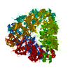 3c3vS S: Starting model for refinement |
|---|---|
| Similar structure data |


-Links
 PDBj
PDBj- Assembly
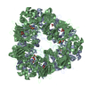
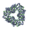
| Deposited unit | 
| ||||||||
|---|---|---|---|---|---|---|---|---|---|
| 1 | 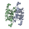
| ||||||||
| Unit cell |
|
-Components
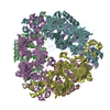
| #1: Protein | Mass: 48178.266 Da / Num. of mol.: 2 / Fragment: UNP residues 43-466 / Mutation: A398E, V416I Source method: isolated from a genetically manipulated source Source: (gene. exp.) Cocos nucifera (coconut palm) / Gene: COS-1 / Production host: Cocos nucifera (coconut palm) / References: UniProt: A0A0R7UCU5, UniProt: A0A222NNM9*PLUS#2: Water | ChemComp-HOH / |  Mass: 18.015 Da / Num. of mol.: 214 / Source method: isolated from a natural source / Formula: H2O Mass: 18.015 Da / Num. of mol.: 214 / Source method: isolated from a natural source / Formula: H2OHas protein modification | Y | |
|---|
-Experimental details
-Experiment
| Experiment | Method: X-RAY DIFFRACTION / Number of used crystals: 1 |
|---|
- Sample preparation
| Crystal | Density Matthews: 1.66 Å3/Da / Density % sol: 32.24 % |
|---|---|
| Crystal grow | Temperature: 288 K / Method: vapor diffusion, hanging drop / pH: 7 / Details: 0.7M NaCl |
-Data collection
| Diffraction | Mean temperature: 100 K |
|---|---|
| Diffraction source | Source: SYNCHROTRON / Site: APS  / Beamline: 22-BM / Wavelength: 1 Å / Beamline: 22-BM / Wavelength: 1 Å |
| Detector | Type: MARMOSAIC 225 mm CCD / Detector: CCD / Date: Aug 12, 2007 |
| Radiation | Protocol: SINGLE WAVELENGTH / Monochromatic (M) / Laue (L): M / Scattering type: x-ray |
| Radiation wavelength | Wavelength: 1 Å / Relative weight: 1 |
| Reflection | Resolution: 1.847→50 Å / Num. obs: 55347 / % possible obs: 95.1 % / Redundancy: 3.6 % / CC1/2: 0.998 / Net I/σ(I): 12.2 |
| Reflection shell | Resolution: 1.85→1.96 Å / Redundancy: 3.7 % / Mean I/σ(I) obs: 2.05 / CC1/2: 0.869 / % possible all: 92.7 |
- Processing
| Software |
| |||||||||||||||||||||||||||||||||||||||||||||||||||||||||||||||||||||||||||||||||||||||||||||||||||||||||||||||||||||||||||||||||||||||||||||||||||
|---|---|---|---|---|---|---|---|---|---|---|---|---|---|---|---|---|---|---|---|---|---|---|---|---|---|---|---|---|---|---|---|---|---|---|---|---|---|---|---|---|---|---|---|---|---|---|---|---|---|---|---|---|---|---|---|---|---|---|---|---|---|---|---|---|---|---|---|---|---|---|---|---|---|---|---|---|---|---|---|---|---|---|---|---|---|---|---|---|---|---|---|---|---|---|---|---|---|---|---|---|---|---|---|---|---|---|---|---|---|---|---|---|---|---|---|---|---|---|---|---|---|---|---|---|---|---|---|---|---|---|---|---|---|---|---|---|---|---|---|---|---|---|---|---|---|---|---|---|
| Refinement | Method to determine structure: MOLECULAR REPLACEMENT Starting model: 3C3V Resolution: 1.847→29.947 Å / SU ML: 0.35 / Cross valid method: FREE R-VALUE / σ(F): 1.91 / Phase error: 36.32
| |||||||||||||||||||||||||||||||||||||||||||||||||||||||||||||||||||||||||||||||||||||||||||||||||||||||||||||||||||||||||||||||||||||||||||||||||||
| Solvent computation | Shrinkage radii: 0.9 Å / VDW probe radii: 1.11 Å | |||||||||||||||||||||||||||||||||||||||||||||||||||||||||||||||||||||||||||||||||||||||||||||||||||||||||||||||||||||||||||||||||||||||||||||||||||
| Refinement step | Cycle: LAST / Resolution: 1.847→29.947 Å
| |||||||||||||||||||||||||||||||||||||||||||||||||||||||||||||||||||||||||||||||||||||||||||||||||||||||||||||||||||||||||||||||||||||||||||||||||||
| Refine LS restraints |
| |||||||||||||||||||||||||||||||||||||||||||||||||||||||||||||||||||||||||||||||||||||||||||||||||||||||||||||||||||||||||||||||||||||||||||||||||||
| LS refinement shell |
|
