 Movie
Movie Controller

[English] 日本語
 Yorodumi
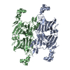
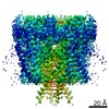
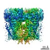
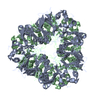
- PDB-1xgf: Backbone Structure of COCOSIN, an 11S storage protein from cocos ... -
+ Open data

- Basic information
| Entry | Database: PDB / ID: 1xgf | ||||||
|---|---|---|---|---|---|---|---|
| Title | Backbone Structure of COCOSIN, an 11S storage protein from cocos nucifera | ||||||
 Components Components | cocosin | ||||||
 Keywords Keywords | Globulin / STORAGE PROTEIN / COCONUT ENDOSPERM / HEXAMER | ||||||
| Biological species |  Cocos nucifera (coconut palm) Cocos nucifera (coconut palm) | ||||||
| Method |  X-RAY DIFFRACTION / MOLECULAR REPLACEMENT / Resolution: 2.61 Å X-RAY DIFFRACTION / MOLECULAR REPLACEMENT / Resolution: 2.61 Å | ||||||
 Authors Authors | Balasundaresan, D. / Ponnuswamy, M.N. | ||||||
 Citation Citation | Journal: TO BE PUBLISHED Title: CRYSTAL STRUCTURE OF COCOSIN FROM COCOS NUCIFERA, A BACKBONE MODEL Authors: Balasundaresan, D. / Ponnuswamy, M.N. #1: Journal: BIOCHIM.BIOPHYS.ACTA / Year: 2002Title: Purification and crystallization of coconut globulin cocosin from cocos nucifera Authors: Balasundaresan, D. / Sugadev, R. / Ponnuswamy, M.N. #2: Journal: Proc.Natl.Acad.Sci.USA / Year: 2003Title: Crystal structure of soybean 11S globulin: Glycinin A3B4 homohexamer Authors: ADACHI, M. / KANAMORI, J. / MASUDA, T. / YAGASAKI, K. / KITAMURA, K. / MIKAMI, B. / UTSUMI, S. | ||||||
| History |
| ||||||
| Remark 400 | COMPOUND THESE ARE SEED STORAGE PROTEINS THAT EXIST AS HEXAMERIC ASSEMBLIES. EACH SUBUNIT IS ...COMPOUND THESE ARE SEED STORAGE PROTEINS THAT EXIST AS HEXAMERIC ASSEMBLIES. EACH SUBUNIT IS COMPOSED OF AN ACIDIC AND A BASIC CHAIN DERIVED FROM A SINGLE PRECURSOR THAT IS LINKED BY A DISULFIDE BOND. MEMBER OF THE 11S FAMILY OF GLOBULINS | ||||||
| Remark 999 | SEQUENCE SINCE NO SEQUENCE INFORMATION IS AVAILABLE,THE RESIDUES HAVE BEEN CHANGED TO UNK AND ONLY ...SEQUENCE SINCE NO SEQUENCE INFORMATION IS AVAILABLE,THE RESIDUES HAVE BEEN CHANGED TO UNK AND ONLY THE BACKBONE COORDINATES ARE GIVEN. THERE ARE FOUR DISORDERED REGIONS IN EACH CHAIN, NAMELY THE RESIDUES FROM 1 TO 6,93 TO 107, 179 TO 199 AND 252 TO 320. |
- Structure visualization
| Structure viewer | Molecule:  MolmilJmol/JSmol MolmilJmol/JSmol |
|---|
- Downloads & links
-Download
| PDBx/mmCIF format | 1xgf.cif.gz | 85.2 KB | Display | PDBx/mmCIF format |
|---|---|---|---|---|
| PDB format | pdb1xgf.ent.gz | 66 KB | Display | PDB format |
| PDBx/mmJSON format | 1xgf.json.gz | Tree view | PDBx/mmJSON format | |
| Others |  Other downloads Other downloads |
-Validation report
| Arichive directory | https://data.pdbj.org/pub/pdb/validation_reports/xg/1xgfftp://data.pdbj.org/pub/pdb/validation_reports/xg/1xgf | HTTPS FTP |
|---|
-Related structure data
| Related structure data |  1od5S S: Starting model for refinement |
|---|---|
| Similar structure data |


-Links
 PDBj
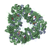
PDBj- Assembly
| Deposited unit | 
| ||||||||
|---|---|---|---|---|---|---|---|---|---|
| 1 | 
| ||||||||
| Unit cell |
| ||||||||
| Details | The biological assembly is a hexamer generated from the molecule in the asymmetric unit by the operation: -Y,X-Y,Z, Y-X,-X,Z, X+2/3,Y+1/3,Z+1/3, -Y+2/3,X-Y+1/3,Z+1/3, Y-X+2/3,-X+1/3,Z+1/3, X+1/3,Y+2/3,Z+2/3, -Y+1/3,X-Y+2/3,Z+2/3, Y-X+1/3,-X+2/3,Z+2/3 |
-Components
| #1: Protein | Mass: 32527.998 Da / Num. of mol.: 2 / Source method: isolated from a natural source / Source: (natural) Cocos nucifera (coconut palm) / Tissue: endosperm |
|---|
-Experimental details
-Experiment
| Experiment | Method: X-RAY DIFFRACTION / Number of used crystals: 1 |
|---|
- Sample preparation
| Crystal | Density Matthews: 4.27 Å3/Da / Density % sol: 70.97 % |
|---|---|
| Crystal grow | Temperature: 293 K / Method: vapor diffusion, hanging drop / pH: 6.7 Details: 20% MPD, Na/K Phosphate buffer containing 7% NaCl, pH 6.7, VAPOR DIFFUSION, HANGING DROP, temperature 293K |
-Data collection
| Diffraction | Mean temperature: 293 K |
|---|---|
| Diffraction source | Source: ROTATING ANODE / Type: RIGAKU RU200 / Wavelength: 1.5418 Å |
| Detector | Type: MARRESEARCH / Detector: IMAGE PLATE / Date: Jan 10, 2002 |
| Radiation | Monochromator: M / Protocol: SINGLE WAVELENGTH / Monochromatic (M) / Laue (L): M / Scattering type: x-ray |
| Radiation wavelength | Wavelength: 1.5418 Å / Relative weight: 1 |
| Reflection | Resolution: 2.6→30 Å / Num. obs: 21429 / % possible obs: 99.8 % / Observed criterion σ(F): 1 |
| Reflection shell | Resolution: 2.6→2.8 Å / Num. unique all: 21429 / % possible all: 93.78 |
- Processing
| Software |
| ||||||||||||||||||||
|---|---|---|---|---|---|---|---|---|---|---|---|---|---|---|---|---|---|---|---|---|---|
| Refinement | Method to determine structure: MOLECULAR REPLACEMENT Starting model: pdb entry 1OD5 Resolution: 2.61→2.8 Å / σ(F): 2 / Stereochemistry target values: Engh & Huber Details: The coordinates contain backbone atoms only and are labelled as UNK since the sequence alignement is not established.There are several abnormal C-N bonds.
| ||||||||||||||||||||
| Refinement step | Cycle: LAST / Resolution: 2.61→2.8 Å
|
