 Movie
Movie Controller


+ Open data

- Basic information
| Entry | Database: PDB / ID: 6az0 | ||||||||||||||||||||||||
|---|---|---|---|---|---|---|---|---|---|---|---|---|---|---|---|---|---|---|---|---|---|---|---|---|---|
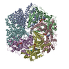
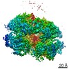
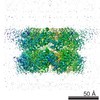
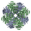
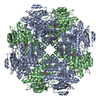
| Title | Mitochondrial ATPase Protease YME1 | ||||||||||||||||||||||||
 Components Components |
| ||||||||||||||||||||||||
 Keywords Keywords | HYDROLASE / Mitochondrial / ATPase / Protease | ||||||||||||||||||||||||
| Function / homology |  Function and homology information Function and homology informationmitochondrial protein quality control / ATP-dependent peptidase activity / mitochondrion organization / metalloendopeptidase activity / mitochondrial inner membrane / ATP hydrolysis activity / ATP binding / metal ion binding Similarity search - Function | ||||||||||||||||||||||||
| Biological species |   | ||||||||||||||||||||||||
| Method | ELECTRON MICROSCOPY / single particle reconstruction / cryo EM / Resolution: 3.4 Å | ||||||||||||||||||||||||
 Authors Authors | Puchades, C. / Rampello, A.J. / Shin, M. / Giuliano, C. / Wiseman, R.L. / Glynn, S.E. / Lander, G.C. | ||||||||||||||||||||||||
| Funding support |  United States, 7items United States, 7items
| ||||||||||||||||||||||||
 Citation Citation | Journal: Science / Year: 2017 Title: Structure of the mitochondrial inner membrane AAA+ protease YME1 gives insight into substrate processing. Authors: Cristina Puchades / Anthony J Rampello / Mia Shin / Christopher J Giuliano / R Luke Wiseman / Steven E Glynn / Gabriel C Lander / Abstract: We present an atomic model of a substrate-bound inner mitochondrial membrane AAA+ quality control protease in yeast, YME1. Our ~3.4-angstrom cryo-electron microscopy structure reveals how the ...We present an atomic model of a substrate-bound inner mitochondrial membrane AAA+ quality control protease in yeast, YME1. Our ~3.4-angstrom cryo-electron microscopy structure reveals how the adenosine triphosphatases (ATPases) form a closed spiral staircase encircling an unfolded substrate, directing it toward the flat, symmetric protease ring. Three coexisting nucleotide states allosterically induce distinct positioning of tyrosines in the central channel, resulting in substrate engagement and translocation to the negatively charged proteolytic chamber. This tight coordination by a network of conserved residues defines a sequential, around-the-ring adenosine triphosphate hydrolysis cycle that results in stepwise substrate translocation. A hingelike linker accommodates the large-scale nucleotide-driven motions of the ATPase spiral relative to the planar proteolytic base. The translocation mechanism is likely conserved for other AAA+ ATPases. | ||||||||||||||||||||||||
| History |
|
- Structure visualization
| Movie |
Movie viewer |
|---|---|
| Structure viewer | Molecule: MolmilJmol/JSmol |
- Downloads & links
-Download
| PDBx/mmCIF format | 6az0.cif.gz | 2 MB | Display | PDBx/mmCIF format |
|---|---|---|---|---|
| PDB format | pdb6az0.ent.gz | 1.8 MB | Display | PDB format |
| PDBx/mmJSON format | 6az0.json.gz | Tree view | PDBx/mmJSON format | |
| Others |  Other downloads Other downloads |
-Validation report
| Arichive directory | https://data.pdbj.org/pub/pdb/validation_reports/az/6az0ftp://data.pdbj.org/pub/pdb/validation_reports/az/6az0 | HTTPS FTP |
|---|
-Related structure data
| Related structure data | 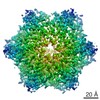 7023MC M: map data used to model this data C: citing same article ( |
|---|---|
| Similar structure data |


-Links
 PDBj
PDBj


- Assembly
| Deposited unit | 
|
|---|---|
| 1 |
|
| Number of models | 5 |
-Components
-Protein / Protein/peptide , 2 types, 7 molecules ABCDEFG
| #1: Protein | Mass: 48025.758 Da / Num. of mol.: 6 Source method: isolated from a genetically manipulated source Details: Walker B mutant Source: (gene. exp.) Strain: RM11-1a / Gene: SCRG_02514 / Production host: #2: Protein/peptide | | Mass: 869.063 Da / Num. of mol.: 1 / Source method: isolated from a natural source / Details: unknown polypeptide / Source: (natural) |
|---|
-Non-polymers , 4 types, 15 molecules 


| #3: Chemical | ChemComp-ATP /  Mass: 507.181 Da / Num. of mol.: 4 / Source method: obtained synthetically / Formula: C10H16N5O13P3 / Comment: ATP, energy-carrying molecule*YM Mass: 507.181 Da / Num. of mol.: 4 / Source method: obtained synthetically / Formula: C10H16N5O13P3 / Comment: ATP, energy-carrying molecule*YM#4: Chemical | ChemComp-ZN /  Mass: 65.409 Da / Num. of mol.: 6 / Source method: obtained synthetically / Formula: Zn Mass: 65.409 Da / Num. of mol.: 6 / Source method: obtained synthetically / Formula: Zn#5: Chemical | ChemComp-MG /  Mass: 24.305 Da / Num. of mol.: 4 / Source method: obtained synthetically / Formula: Mg Mass: 24.305 Da / Num. of mol.: 4 / Source method: obtained synthetically / Formula: Mg#6: Chemical | ChemComp-ADP / |  Mass: 427.201 Da / Num. of mol.: 1 / Source method: obtained synthetically / Formula: C10H15N5O10P2 / Comment: ADP, energy-carrying molecule*YM Mass: 427.201 Da / Num. of mol.: 1 / Source method: obtained synthetically / Formula: C10H15N5O10P2 / Comment: ADP, energy-carrying molecule*YM |
|---|
-Experimental details
-Experiment
| Experiment | Method: ELECTRON MICROSCOPY |
|---|---|
| EM experiment | Aggregation state: PARTICLE / 3D reconstruction method: single particle reconstruction |
- Sample preparation
| Component | Name: YME1 ATPase Protease Walker B mutant bound to substrate Type: COMPLEX Details: YME1 Walker B mutant was recombinantly expressed in E. coli, solved in presence of ATP with unknown bound substrate. Entity ID: #1-#2 / Source: RECOMBINANT | |||||||||||||||||||||||||||||||||||
|---|---|---|---|---|---|---|---|---|---|---|---|---|---|---|---|---|---|---|---|---|---|---|---|---|---|---|---|---|---|---|---|---|---|---|---|---|
| Molecular weight | Value: 0.295 MDa / Experimental value: NO | |||||||||||||||||||||||||||||||||||
| Source (natural) | Organism: | |||||||||||||||||||||||||||||||||||
| Source (recombinant) | Organism: | |||||||||||||||||||||||||||||||||||
| Buffer solution | pH: 8 | |||||||||||||||||||||||||||||||||||
| Buffer component |
| |||||||||||||||||||||||||||||||||||
| Specimen | Embedding applied: NO / Shadowing applied: NO / Staining applied: NO / Vitrification applied: YES | |||||||||||||||||||||||||||||||||||
| Specimen support | Details: Gatan Solarus Plasma Cleaner / Grid material: GOLD / Grid mesh size: 300 divisions/in. / Grid type: Quantifoil R1.2/1.3 | |||||||||||||||||||||||||||||||||||
| Vitrification | Instrument: FEI VITROBOT MARK IV / Cryogen name: ETHANE / Humidity: 100 % / Chamber temperature: 298 K |
- Electron microscopy imaging
| Experimental equipment |  Model: Titan Krios / Image courtesy: FEI Company |
|---|---|
| Microscopy | Model: FEI TITAN KRIOS / Details: Data collected using Leginon |
| Electron gun | Electron source:  FIELD EMISSION GUN / Accelerating voltage: 300 kV / Illumination mode: FLOOD BEAM FIELD EMISSION GUN / Accelerating voltage: 300 kV / Illumination mode: FLOOD BEAM |
| Electron lens | Mode: BRIGHT FIELD / Nominal magnification: 29000 X / Calibrated magnification: 51500 X / Nominal defocus max: 2500 nm / Nominal defocus min: 1200 nm / Calibrated defocus min: 1200 nm / Calibrated defocus max: 2500 nm / Cs: 2.7 mm / C2 aperture diameter: 100 µm / Alignment procedure: COMA FREE |
| Specimen holder | Cryogen: NITROGEN / Specimen holder model: FEI TITAN KRIOS AUTOGRID HOLDER / Temperature (max): 70 K / Temperature (min): 70 K |
| Image recording | Average exposure time: 7 sec. / Electron dose: 60 e/Å2 / Detector mode: COUNTING / Film or detector model: GATAN K2 SUMMIT (4k x 4k) / Num. of grids imaged: 3 / Num. of real images: 6098 Details: Images collected in counting mode at 5 frames per second. |
| Image scans | Sampling size: 5 µm / Width: 3710 / Height: 3838 / Movie frames/image: 35 / Used frames/image: 1-35 |
- Processing
| Software | Name: PHENIX / Version: 1.10.1_2155: / Classification: refinement | |||||||||||||||||||||||||||||||||||||||||||||
|---|---|---|---|---|---|---|---|---|---|---|---|---|---|---|---|---|---|---|---|---|---|---|---|---|---|---|---|---|---|---|---|---|---|---|---|---|---|---|---|---|---|---|---|---|---|---|
| EM software |
| |||||||||||||||||||||||||||||||||||||||||||||
| CTF correction | Type: PHASE FLIPPING ONLY | |||||||||||||||||||||||||||||||||||||||||||||
| Particle selection | Num. of particles selected: 2285499 Details: Particles picked automatically with FindEM template picking | |||||||||||||||||||||||||||||||||||||||||||||
| Symmetry | Point symmetry: C1 (asymmetric) | |||||||||||||||||||||||||||||||||||||||||||||
| 3D reconstruction | Resolution: 3.4 Å / Resolution method: FSC 0.143 CUT-OFF / Num. of particles: 62917 / Algorithm: BACK PROJECTION / Symmetry type: POINT | |||||||||||||||||||||||||||||||||||||||||||||
| Atomic model building | B value: 100 / Protocol: AB INITIO MODEL / Space: REAL | |||||||||||||||||||||||||||||||||||||||||||||
| Refine LS restraints |
|
