Movie
Movie Controller

[English] 日本語
 Yorodumi
- EMDB-4626: ClpB (DWB and K476C mutant) bound to casein in presence of ATPgam... -
+ Open data

- Basic information
| Entry | Database: EMDB / ID: EMD-4626 | ||||||||||||
|---|---|---|---|---|---|---|---|---|---|---|---|---|---|
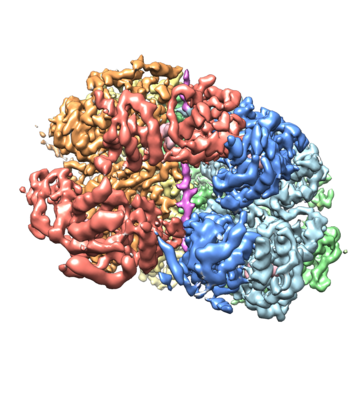
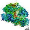
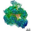
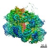
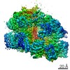
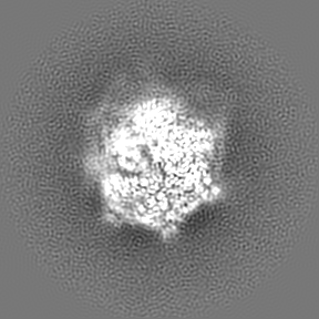
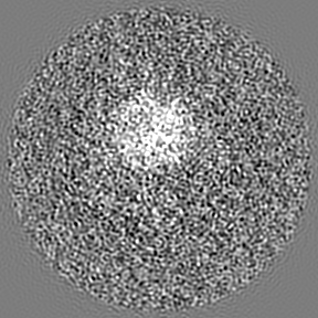
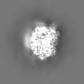
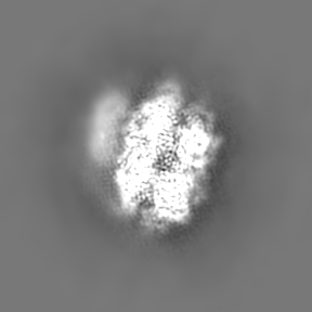
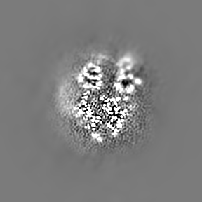
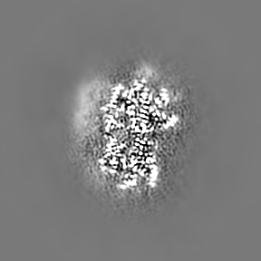
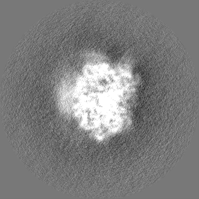
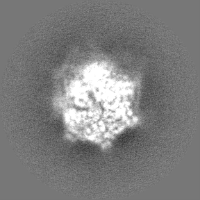
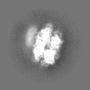
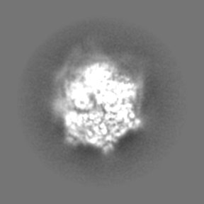
| Title | ClpB (DWB and K476C mutant) bound to casein in presence of ATPgammaS - state KC-2A | ||||||||||||
 Map data Map data | ClpB (DWB and K476C mutant) bound to a casein substrate in presence of ATPgammaS - State KC2A | ||||||||||||
 Sample Sample |
| ||||||||||||
 Keywords Keywords | disaggregase / proteostasis / AAA / CHAPERONE | ||||||||||||
| Function / homology |  Function and homology information Function and homology informationcellular response to heat / response to heat / protein refolding / ATP hydrolysis activity / ATP binding / membrane / identical protein binding / cytoplasm / cytosol Similarity search - Function | ||||||||||||
| Biological species |   | ||||||||||||
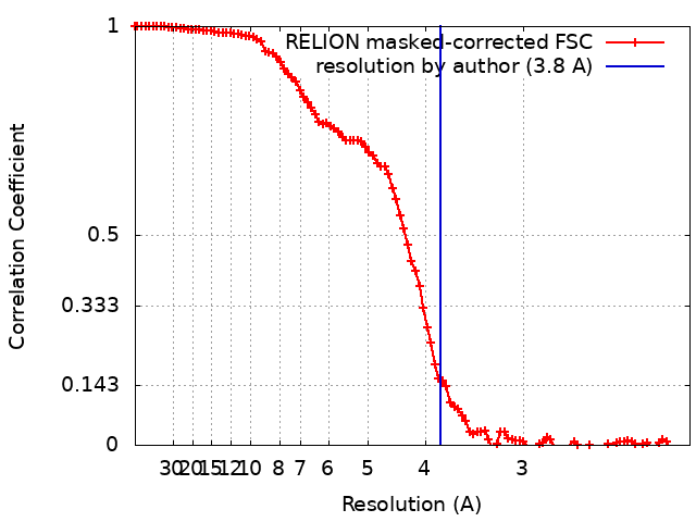
| Method | single particle reconstruction / cryo EM / Resolution: 3.8 Å | ||||||||||||
 Authors Authors | Deville C / Saibil HR | ||||||||||||
| Funding support |  United Kingdom, 3 items United Kingdom, 3 items
| ||||||||||||
 Citation Citation | Journal: Cell Rep / Year: 2019 Title: Two-Step Activation Mechanism of the ClpB Disaggregase for Sequential Substrate Threading by the Main ATPase Motor. Authors: Célia Deville / Kamila Franke / Axel Mogk / Bernd Bukau / Helen R Saibil /  Abstract: AAA+ proteins form asymmetric hexameric rings that hydrolyze ATP and thread substrate proteins through a central channel via mobile substrate-binding pore loops. Understanding how ATPase and ...AAA+ proteins form asymmetric hexameric rings that hydrolyze ATP and thread substrate proteins through a central channel via mobile substrate-binding pore loops. Understanding how ATPase and threading activities are regulated and intertwined is key to understanding the AAA+ protein mechanism. We studied the disaggregase ClpB, which contains tandem ATPase domains (AAA1, AAA2) and shifts between low and high ATPase and threading activities. Coiled-coil M-domains repress ClpB activity by encircling the AAA1 ring. Here, we determine the mechanism of ClpB activation by comparing ATPase mechanisms and cryo-EM structures of ClpB wild-type and a constitutively active ClpB M-domain mutant. We show that ClpB activation reduces ATPase cooperativity and induces a sequential mode of ATP hydrolysis in the AAA2 ring, the main ATPase motor. AAA1 and AAA2 rings do not work synchronously but in alternating cycles. This ensures high grip, enabling substrate threading via a processive, rope-climbing mechanism. | ||||||||||||
| History |
|
- Structure visualization
| Movie |
Movie viewer |
|---|---|
| Structure viewer | EM map: SurfViewMolmilJmol/JSmol |
| Supplemental images |
- Downloads & links
-EMDB archive
| Map data | emd_4626.map.gz | 85.4 MB | EMDB map data format | |
|---|---|---|---|---|
| Header (meta data) | emd-4626-v30.xmlemd-4626.xml | 25.6 KB 25.6 KB | Display Display | EMDB header |
| FSC (resolution estimation) | emd_4626_fsc.xml | 10.3 KB | Display | FSC data file |
| Images |  emd_4626.png emd_4626.png | 163.7 KB | ||
| Filedesc metadata | emd-4626.cif.gz | 7.4 KB | ||
| Others | emd_4626_additional.map.gzemd_4626_half_map_1.map.gzemd_4626_half_map_2.map.gz | 57.7 MB 71.3 MB 29.7 MB | ||
| Archive directory |  http://ftp.pdbj.org/pub/emdb/structures/EMD-4626ftp://ftp.pdbj.org/pub/emdb/structures/EMD-4626 http://ftp.pdbj.org/pub/emdb/structures/EMD-4626ftp://ftp.pdbj.org/pub/emdb/structures/EMD-4626 | HTTPS FTP |
-Related structure data
| Related structure data |  6qs7MC 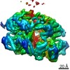 4621C 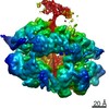 4622C 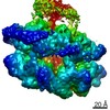 4623C 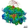 4624C 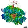 4625C 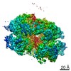 4627C 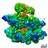 4940C  4941C 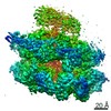 4942C  6qs4C  6qs6C  6qs8C  6rn2C 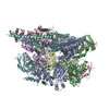 6rn3C 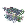 6rn4C C: citing same article ( M: atomic model generated by this map |
|---|---|
| Similar structure data |
-Links
| EMDB pages | EMDB (EBI/PDBe) / EMDataResource |
|---|---|
| Related items in Molecule of the Month |


-Map
| File | Download / File: emd_4626.map.gz / Format: CCP4 / Size: 91.1 MB / Type: IMAGE STORED AS FLOATING POINT NUMBER (4 BYTES) | ||||||||||||||||||||||||||||||||||||||||||||||||||||||||||||||||||||
|---|---|---|---|---|---|---|---|---|---|---|---|---|---|---|---|---|---|---|---|---|---|---|---|---|---|---|---|---|---|---|---|---|---|---|---|---|---|---|---|---|---|---|---|---|---|---|---|---|---|---|---|---|---|---|---|---|---|---|---|---|---|---|---|---|---|---|---|---|---|
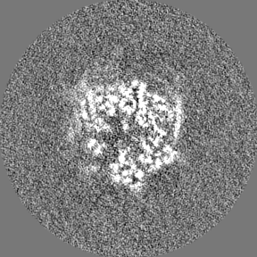
| Annotation | ClpB (DWB and K476C mutant) bound to a casein substrate in presence of ATPgammaS - State KC2A | ||||||||||||||||||||||||||||||||||||||||||||||||||||||||||||||||||||
| Projections & slices | Image control
Images are generated by Spider. | ||||||||||||||||||||||||||||||||||||||||||||||||||||||||||||||||||||
| Voxel size | X=Y=Z: 1.05 Å | ||||||||||||||||||||||||||||||||||||||||||||||||||||||||||||||||||||
| Density |
| ||||||||||||||||||||||||||||||||||||||||||||||||||||||||||||||||||||
| Symmetry | Space group: 1 | ||||||||||||||||||||||||||||||||||||||||||||||||||||||||||||||||||||
| Details | EMDB XML:
CCP4 map header:
| ||||||||||||||||||||||||||||||||||||||||||||||||||||||||||||||||||||
 Z (Sec.)
 Y (Row.)
 X (Col.)


-Supplemental data
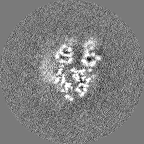
-Additional map: map filtered according to local resolution
| File | emd_4626_additional.map | ||||||||||||
|---|---|---|---|---|---|---|---|---|---|---|---|---|---|
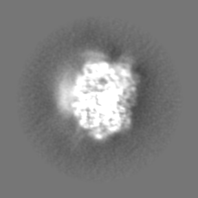
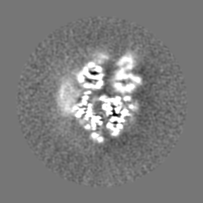
| Annotation | map filtered according to local resolution | ||||||||||||
| Projections & Slices |
| ||||||||||||
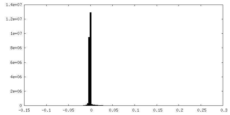
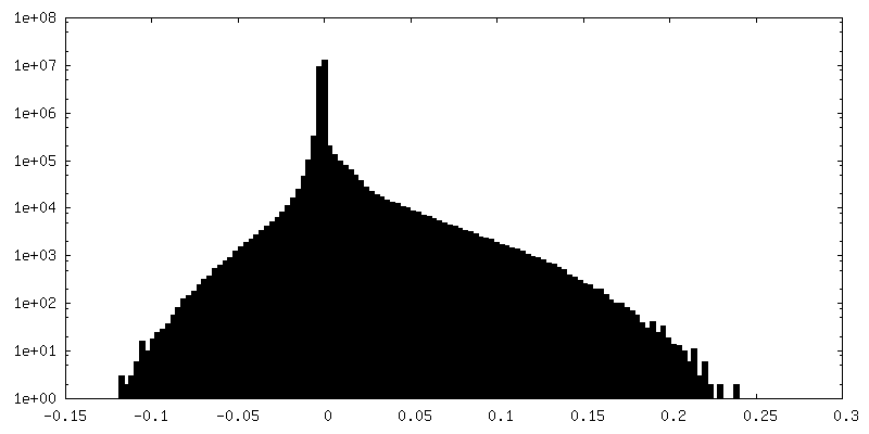
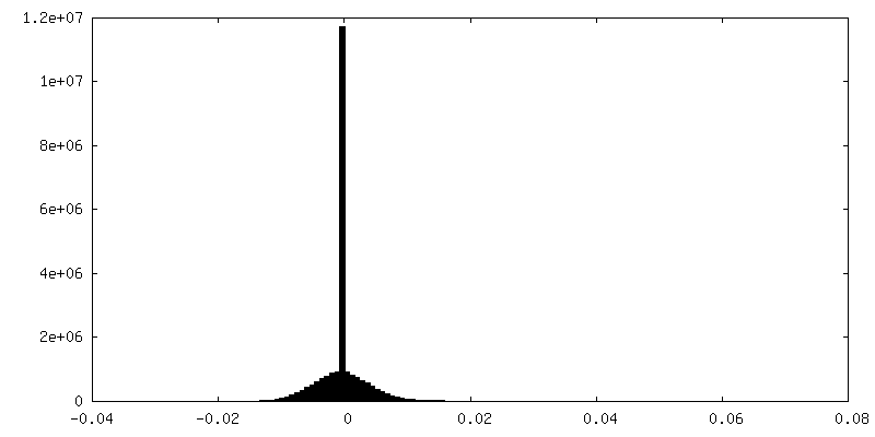
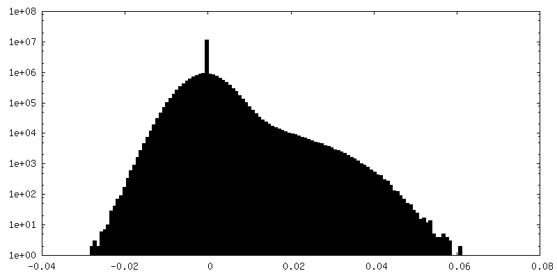
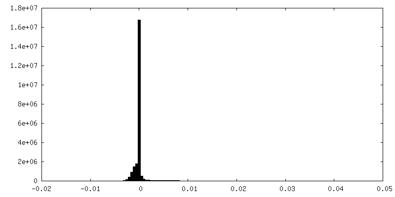
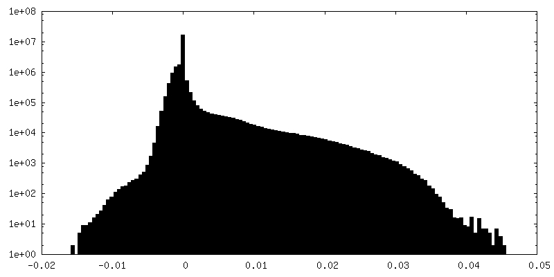
| Density Histograms |


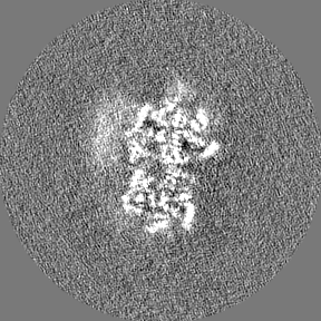
-Half map: Half map 1
| File | emd_4626_half_map_1.map | ||||||||||||
|---|---|---|---|---|---|---|---|---|---|---|---|---|---|
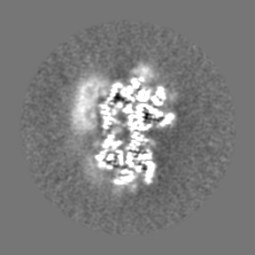
| Annotation | Half map 1 | ||||||||||||
| Projections & Slices |
| ||||||||||||
| Density Histograms |

-Half map: Half map 2
| File | emd_4626_half_map_2.map | ||||||||||||
|---|---|---|---|---|---|---|---|---|---|---|---|---|---|
| Annotation | Half map 2 | ||||||||||||
| Projections & Slices |
| ||||||||||||
| Density Histograms |
- Sample components
-Entire : ClpB-DWB-K476C bound to casein in presence of ATPgammaS
| Entire | Name: ClpB-DWB-K476C bound to casein in presence of ATPgammaS |
|---|---|
| Components |
|
-Supramolecule #1: ClpB-DWB-K476C bound to casein in presence of ATPgammaS
| Supramolecule | Name: ClpB-DWB-K476C bound to casein in presence of ATPgammaS type: complex / ID: 1 / Parent: 0 / Macromolecule list: #1-#2 |
|---|---|
| Molecular weight | Theoretical: 570 KDa |
-Supramolecule #2: ClpB-DWB-K476C
| Supramolecule | Name: ClpB-DWB-K476C / type: complex / ID: 2 / Parent: 1 / Macromolecule list: #1 |
|---|---|
| Source (natural) | Organism: |
-Supramolecule #3: casein
| Supramolecule | Name: casein / type: complex / ID: 3 / Parent: 1 / Macromolecule list: #2 |
|---|---|
| Source (natural) | Organism: |
-Macromolecule #1: Chaperone protein ClpB
| Macromolecule | Name: Chaperone protein ClpB / type: protein_or_peptide / ID: 1 / Number of copies: 6 / Enantiomer: LEVO |
|---|---|
| Source (natural) | Organism: |
| Molecular weight | Theoretical: 95.708133 KDa |
| Recombinant expression | Organism: |
| Sequence | String: MRLDRLTNKF QLALADAQSL ALGHDNQFIE PLHLMSALLN QEGGSVSPLL TSAGINAGQL RTDINQALNR LPQVEGTGGD VQPSQDLVR VLNLCDKLAQ KRGDNFISSE LFVLAALESR GTLADILKAA GATTANITQA IEQMRGGESV NDQGAEDQRQ A LKKYTIDL ...String: MRLDRLTNKF QLALADAQSL ALGHDNQFIE PLHLMSALLN QEGGSVSPLL TSAGINAGQL RTDINQALNR LPQVEGTGGD VQPSQDLVR VLNLCDKLAQ KRGDNFISSE LFVLAALESR GTLADILKAA GATTANITQA IEQMRGGESV NDQGAEDQRQ A LKKYTIDL TERAEQGKLD PVIGRDEEIR RTIQVLQRRT KNNPVLIGEP GVGKTAIVEG LAQRIINGEV PEGLKGRRVL AL DMGALVA GAKYRGEFEE RLKGVLNDLA KQEGNVILFI DALHTMVGAG KADGAMDAGN MLKPALARGE LHCVGATTLD EYR QYIEKD AALERRFQKV FVAEPSVEDT IAILRGLKER YELHHHVQIT DPAIVAAATL SHRYIADRQL PDKAIDLIDE AASS IRMQI DSKPEELDRL DRRIIQLKLE QQALMKESDE ASKKRLDMLN EELSDKERQY SELEEEWKAE KASLSGTQTI CAELE QAKI AIEQARRVGD LARMSELQYG KIPELEKQLE AATQLEGKTM RLLRNKVTDA EIAEVLARWT GIPVSRMMES EREKLL RME QELHHRVIGQ NEAVDAVSNA IRRSRAGLAD PNRPIGSFLF LGPTGVGKTE LCKALANFMF DSDEAMVRID MSEFMEK HS VSRLVGAPPG YVGYEEGGYL TEAVRRRPYS VILLDAVEKA HPDVFNILLQ VLDDGRLTDG QGRTVDFRNT VVIMTSNL G SDLIQERFGE LDYAHMKELV LGVVSHNFRP EFINRIDEVV VFHPLGEQHI ASIAQIQLKR LYKRLEERGY EIHISDEAL KLLSENGYDP VYGARPLKRA IQQQIENPLA QQILSGELVP GKVIRLEVNE DRIVAVQH UniProtKB: Chaperone protein ClpB |
-Macromolecule #2: casein
| Macromolecule | Name: casein / type: protein_or_peptide / ID: 2 / Number of copies: 1 / Enantiomer: LEVO |
|---|---|
| Source (natural) | Organism: |
| Molecular weight | Theoretical: 2.060531 KDa |
| Sequence | String: (UNK)(UNK)(UNK)(UNK)(UNK)(UNK)(UNK)(UNK)(UNK)(UNK) (UNK)(UNK)(UNK)(UNK)(UNK)(UNK) (UNK)(UNK)(UNK) (UNK)(UNK)(UNK)(UNK)(UNK) |
-Macromolecule #3: PHOSPHOTHIOPHOSPHORIC ACID-ADENYLATE ESTER
| Macromolecule | Name: PHOSPHOTHIOPHOSPHORIC ACID-ADENYLATE ESTER / type: ligand / ID: 3 / Number of copies: 10 / Formula: AGS |
|---|---|
| Molecular weight | Theoretical: 523.247 Da |
| Chemical component information |  ChemComp-AGS: |
-Macromolecule #4: MAGNESIUM ION
| Macromolecule | Name: MAGNESIUM ION / type: ligand / ID: 4 / Number of copies: 8 / Formula: MG |
|---|---|
| Molecular weight | Theoretical: 24.305 Da |
-Macromolecule #5: ADENOSINE-5'-DIPHOSPHATE
| Macromolecule | Name: ADENOSINE-5'-DIPHOSPHATE / type: ligand / ID: 5 / Number of copies: 1 / Formula: ADP |
|---|---|
| Molecular weight | Theoretical: 427.201 Da |
| Chemical component information |  ChemComp-ADP: |
-Experimental details
-Structure determination
| Method | cryo EM |
|---|---|
 Processing Processing | single particle reconstruction |
| Aggregation state | particle |
-Sample preparation
| Concentration | 1.6 mg/mL | ||||||||||||||||||
|---|---|---|---|---|---|---|---|---|---|---|---|---|---|---|---|---|---|---|---|
| Buffer | pH: 7.5 Component:
| ||||||||||||||||||
| Grid | Model: Quantifoil, UltrAuFoil / Material: GOLD / Mesh: 300 / Support film - Material: GRAPHENE OXIDE / Support film - topology: CONTINUOUS / Pretreatment - Type: GLOW DISCHARGE / Pretreatment - Time: 60 sec. / Pretreatment - Atmosphere: AIR | ||||||||||||||||||
| Vitrification | Cryogen name: ETHANE / Chamber humidity: 100 % / Chamber temperature: 295 K / Instrument: FEI VITROBOT MARK IV |
- Electron microscopy
| Microscope | FEI TITAN KRIOS |
|---|---|
| Specialist optics | Energy filter - Name: GIF Bioquantum |
| Image recording | Film or detector model: GATAN K2 SUMMIT (4k x 4k) / Detector mode: COUNTING / Number grids imaged: 1 / Number real images: 6060 / Average electron dose: 1.0 e/Å2 |
| Electron beam | Acceleration voltage: 300 kV / Electron source:  FIELD EMISSION GUN FIELD EMISSION GUN |
| Electron optics | C2 aperture diameter: 70.0 µm / Illumination mode: FLOOD BEAM / Imaging mode: BRIGHT FIELD / Cs: 2.7 mm / Nominal defocus max: 3.5 µm / Nominal defocus min: 1.5 µm |
| Sample stage | Specimen holder model: FEI TITAN KRIOS AUTOGRID HOLDER / Cooling holder cryogen: NITROGEN |
| Experimental equipment |  Model: Titan Krios / Image courtesy: FEI Company |