+ Open data
- Basic information
| Entry | Database: PDB / ID: 6nyy | ||||||
|---|---|---|---|---|---|---|---|
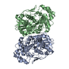
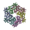
| Title | human m-AAA protease AFG3L2, substrate-bound | ||||||
 Components Components |
| ||||||
 Keywords Keywords | TRANSLOCASE / AAA+ / ATPase / protease / mitochondria / protein quality control / neurodegeneration / inner membrane / AFG3L2 / m/AAA protease | ||||||
| Function / homology |  Function and homology information Function and homology informationcellular response to glutathione / m-AAA complex / mitochondrial protein processing / regulation of calcium import into the mitochondrion / mitochondrial protein quality control / Processing of SMDT1 / mitochondrial calcium ion homeostasis / membrane protein proteolysis / calcium import into the mitochondrion / Hydrolases; Acting on acid anhydrides ...cellular response to glutathione / m-AAA complex / mitochondrial protein processing / regulation of calcium import into the mitochondrion / mitochondrial protein quality control / Processing of SMDT1 / mitochondrial calcium ion homeostasis / membrane protein proteolysis / calcium import into the mitochondrion / Hydrolases; Acting on acid anhydrides / cristae formation / righting reflex / nerve development / muscle cell development / Hydrolases; Acting on peptide bonds (peptidases); Metalloendopeptidases / mitochondrial fusion / ATP-dependent peptidase activity / neuromuscular junction development / protein autoprocessing / regulation of multicellular organism growth / myelination / Mitochondrial protein degradation / axonogenesis / protein maturation / protein catabolic process / metalloendopeptidase activity / protein processing / metallopeptidase activity / unfolded protein binding / mitochondrial inner membrane / ATP hydrolysis activity / mitochondrion / proteolysis / zinc ion binding / ATP binding Similarity search - Function | ||||||
| Biological species |  Homo sapiens (human) Homo sapiens (human) | ||||||
| Method | ELECTRON MICROSCOPY / single particle reconstruction / cryo EM / Resolution: 3 Å | ||||||
 Authors Authors | Lander, G.C. / Puchades, C. | ||||||
| Funding support |  United States, 1items United States, 1items
| ||||||
 Citation Citation |  Journal: Mol Cell / Year: 2019 Journal: Mol Cell / Year: 2019Title: Unique Structural Features of the Mitochondrial AAA+ Protease AFG3L2 Reveal the Molecular Basis for Activity in Health and Disease. Authors: Cristina Puchades / Bojian Ding / Albert Song / R Luke Wiseman / Gabriel C Lander / Steven E Glynn /  Abstract: Mitochondrial AAA+ quality-control proteases regulate diverse aspects of mitochondrial biology through specialized protein degradation, but the underlying mechanisms of these enzymes remain poorly ...Mitochondrial AAA+ quality-control proteases regulate diverse aspects of mitochondrial biology through specialized protein degradation, but the underlying mechanisms of these enzymes remain poorly defined. The mitochondrial AAA+ protease AFG3L2 is of particular interest, as genetic mutations localized throughout AFG3L2 are linked to diverse neurodegenerative disorders. However, a lack of structural data has limited our understanding of how mutations impact enzymatic function. Here, we used cryoelectron microscopy (cryo-EM) to determine a substrate-bound structure of the catalytic core of human AFG3L2. This structure identifies multiple specialized structural features that integrate with conserved motifs required for ATP-dependent translocation to unfold and degrade targeted proteins. Many disease-relevant mutations localize to these unique structural features of AFG3L2 and distinctly influence its activity and stability. Our results provide a molecular basis for neurological phenotypes associated with different AFG3L2 mutations and establish a structural framework to understand how different members of the AAA+ superfamily achieve specialized biological functions. | ||||||
| History |
|
- Structure visualization
| Movie |
 Movie viewer Movie viewer |
|---|---|
| Structure viewer | Molecule:  Molmil Molmil Jmol/JSmol Jmol/JSmol |
- Downloads & links
- Download
| PDBx/mmCIF format |  6nyy.cif.gz 6nyy.cif.gz | 453.4 KB | Display |  PDBx/mmCIF format PDBx/mmCIF format |
|---|---|---|---|---|
| PDB format |  pdb6nyy.ent.gz pdb6nyy.ent.gz | 351.9 KB | Display |  PDB format PDB format |
| PDBx/mmJSON format |  6nyy.json.gz 6nyy.json.gz | Tree view |  PDBx/mmJSON format PDBx/mmJSON format | |
| Others |  Other downloads Other downloads |
-Validation report
| Arichive directory |  https://data.pdbj.org/pub/pdb/validation_reports/ny/6nyy https://data.pdbj.org/pub/pdb/validation_reports/ny/6nyy ftp://data.pdbj.org/pub/pdb/validation_reports/ny/6nyy ftp://data.pdbj.org/pub/pdb/validation_reports/ny/6nyy | HTTPS FTP |
|---|
-Related structure data
| Related structure data |  0552MC M: map data used to model this data C: citing same article ( |
|---|---|
| Similar structure data |
- Links
Links
- Assembly
| Deposited unit | 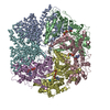
|
|---|---|
| 1 |
|
- Components
-Protein / Protein/peptide , 2 types, 10 molecules ABCDEFGHIJ
| #1: Protein | Mass: 58331.629 Da / Num. of mol.: 6 Source method: isolated from a genetically manipulated source Source: (gene. exp.)  Homo sapiens (human) / Gene: AFG3L2 / Production host: Homo sapiens (human) / Gene: AFG3L2 / Production host:  References: UniProt: Q9Y4W6, Hydrolases; Acting on peptide bonds (peptidases); Metalloendopeptidases #2: Protein/peptide | Mass: 799.871 Da / Num. of mol.: 4 Source method: isolated from a genetically manipulated source Source: (gene. exp.)  Homo sapiens (human) / Production host: Homo sapiens (human) / Production host:  |
|---|
-Non-polymers , 4 types, 13 molecules 


| #3: Chemical | ChemComp-ZN / #4: Chemical | #5: Chemical | #6: Chemical | ChemComp-ADP / | |
|---|
-Experimental details
-Experiment
| Experiment | Method: ELECTRON MICROSCOPY |
|---|---|
| EM experiment | Aggregation state: PARTICLE / 3D reconstruction method: single particle reconstruction |
- Sample preparation
| Component | Name: AFG3L2 / Type: ORGANELLE OR CELLULAR COMPONENT Details: Soluble domains of the AFG3L2, comprising the AAA+ ATPase and protease domains. Entity ID: #1-#2 / Source: RECOMBINANT | ||||||||||||||||||||||||||||||
|---|---|---|---|---|---|---|---|---|---|---|---|---|---|---|---|---|---|---|---|---|---|---|---|---|---|---|---|---|---|---|---|
| Molecular weight | Value: .360 MDa / Experimental value: NO | ||||||||||||||||||||||||||||||
| Source (natural) | Organism:  Homo sapiens (human) Homo sapiens (human) | ||||||||||||||||||||||||||||||
| Source (recombinant) | Organism:  | ||||||||||||||||||||||||||||||
| Buffer solution | pH: 8 Details: DTT and ATP were added fresh before sample preparation | ||||||||||||||||||||||||||||||
| Buffer component |
| ||||||||||||||||||||||||||||||
| Specimen | Conc.: 2 mg/ml / Embedding applied: NO / Shadowing applied: NO / Staining applied: NO / Vitrification applied: YES | ||||||||||||||||||||||||||||||
| Specimen support | Details: Grids were plasma cleaned using a Solarus plasma cleaner (Gatan, Inc.). Grid material: GOLD / Grid mesh size: 300 divisions/in. / Grid type: Quantifoil, UltrAuFoil, R1.2/1.3 | ||||||||||||||||||||||||||||||
| Vitrification | Instrument: FEI VITROBOT MARK III / Cryogen name: ETHANE / Humidity: 100 % / Chamber temperature: 278 K Details: Sample was blotted for 3 seconds using Whatman No. 1 filter paper immediately prio to plunge-freezing. |
- Electron microscopy imaging
| Experimental equipment |  Model: Talos Arctica / Image courtesy: FEI Company |
|---|---|
| Microscopy | Model: FEI TECNAI ARCTICA |
| Electron gun | Electron source:  FIELD EMISSION GUN / Accelerating voltage: 200 kV / Illumination mode: FLOOD BEAM FIELD EMISSION GUN / Accelerating voltage: 200 kV / Illumination mode: FLOOD BEAM |
| Electron lens | Mode: BRIGHT FIELD / Nominal magnification: 36000 X / Calibrated magnification: 43478 X / Nominal defocus max: 18000 nm / Nominal defocus min: 8000 nm / Cs: 2.7 mm / C2 aperture diameter: 70 µm / Alignment procedure: COMA FREE |
| Specimen holder | Cryogen: NITROGEN / Specimen holder model: FEI TITAN KRIOS AUTOGRID HOLDER / Temperature (max): 100 K / Temperature (min): 100 K / Residual tilt: 0.07 mradians |
| Image recording | Average exposure time: 7 sec. / Electron dose: 40 e/Å2 / Detector mode: COUNTING / Film or detector model: GATAN K2 SUMMIT (4k x 4k) / Num. of grids imaged: 5 / Num. of real images: 5707 / Details: Data collected with Leginon |
| Image scans | Sampling size: 5 µm / Width: 3710 / Height: 3838 / Movie frames/image: 30 / Used frames/image: 1-30 |
- Processing
| Software | Name: PHENIX / Version: 1.13_2998: / Classification: refinement | ||||||||||||||||||||||||||||||||||||||||||||||||||
|---|---|---|---|---|---|---|---|---|---|---|---|---|---|---|---|---|---|---|---|---|---|---|---|---|---|---|---|---|---|---|---|---|---|---|---|---|---|---|---|---|---|---|---|---|---|---|---|---|---|---|---|
| EM software |
| ||||||||||||||||||||||||||||||||||||||||||||||||||
| CTF correction | Details: CTF estimated with CTFFIND 4, local CTF estimated with Type: PHASE FLIPPING AND AMPLITUDE CORRECTION | ||||||||||||||||||||||||||||||||||||||||||||||||||
| Particle selection | Num. of particles selected: 4541491 / Details: Picked with FindEM | ||||||||||||||||||||||||||||||||||||||||||||||||||
| Symmetry | Point symmetry: C1 (asymmetric) | ||||||||||||||||||||||||||||||||||||||||||||||||||
| 3D reconstruction | Resolution: 3 Å / Resolution method: FSC 0.143 CUT-OFF / Num. of particles: 1129437 / Algorithm: BACK PROJECTION Details: Multi-body refinement was used to generate 3 different reconstructions that were stitched together Symmetry type: POINT | ||||||||||||||||||||||||||||||||||||||||||||||||||
| Atomic model building | B value: 85 / Protocol: RIGID BODY FIT / Space: REAL Details: The sequence was threaded onto the PDB ID 6AZ0 and built into the EM density with Coot. | ||||||||||||||||||||||||||||||||||||||||||||||||||
| Atomic model building | PDB-ID: 6AZ0 Accession code: 6AZ0 / Source name: PDB / Type: experimental model |
 Movie
Movie Controller





 PDBj
PDBj


