 Movie
Movie Controller


+ Open data

- Basic information
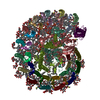
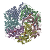
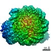
| Entry | Database: EMDB / ID: EMD-11081 | |||||||||
|---|---|---|---|---|---|---|---|---|---|---|
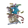
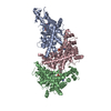
| Title | RC-LH1(14)-W complex from Rhodopseudomonas palustris | |||||||||
 Map data Map data | ||||||||||
 Sample Sample |
| |||||||||
 Keywords Keywords | Reaction center / Light harvesting / Protein W / Quinone / PHOTOSYNTHESIS | |||||||||
| Function / homology |  Function and homology information Function and homology informationorganelle inner membrane / plasma membrane-derived chromatophore membrane / plasma membrane light-harvesting complex / bacteriochlorophyll binding / photosynthetic electron transport in photosystem II / photosynthesis, light reaction / metal ion binding / plasma membrane Similarity search - Function | |||||||||
| Biological species |  Rhodopseudomonas palustris CGA009 (phototrophic) / Rhodopseudomonas palustris (strain ATCC BAA-98 / CGA009) (phototrophic) Rhodopseudomonas palustris CGA009 (phototrophic) / Rhodopseudomonas palustris (strain ATCC BAA-98 / CGA009) (phototrophic) | |||||||||
| Method | single particle reconstruction / cryo EM / Resolution: 2.65 Å | |||||||||
 Authors Authors | Swainsbury DJK / Qian P | |||||||||
| Funding support |  United Kingdom, 1 items United Kingdom, 1 items
| |||||||||
 Citation Citation | Journal: Sci Adv / Year: 2021 Title: Structures of RC-LH1 complexes with open or closed quinone channels. Authors: David J K Swainsbury / Pu Qian / Philip J Jackson / Kaitlyn M Faries / Dariusz M Niedzwiedzki / Elizabeth C Martin / David A Farmer / Lorna A Malone / Rebecca F Thompson / Neil A Ranson / ...Authors: David J K Swainsbury / Pu Qian / Philip J Jackson / Kaitlyn M Faries / Dariusz M Niedzwiedzki / Elizabeth C Martin / David A Farmer / Lorna A Malone / Rebecca F Thompson / Neil A Ranson / Daniel P Canniffe / Mark J Dickman / Dewey Holten / Christine Kirmaier / Andrew Hitchcock / C Neil Hunter /   Abstract: The reaction-center light-harvesting complex 1 (RC-LH1) is the core photosynthetic component in purple phototrophic bacteria. We present two cryo-electron microscopy structures of RC-LH1 complexes ...The reaction-center light-harvesting complex 1 (RC-LH1) is the core photosynthetic component in purple phototrophic bacteria. We present two cryo-electron microscopy structures of RC-LH1 complexes from A 2.65-Å resolution structure of the RC-LH1-W complex consists of an open 14-subunit LH1 ring surrounding the RC interrupted by protein-W, whereas the complex without protein-W at 2.80-Å resolution comprises an RC completely encircled by a closed, 16-subunit LH1 ring. Comparison of these structures provides insights into quinone dynamics within RC-LH1 complexes, including a previously unidentified conformational change upon quinone binding at the RC Q site, and the locations of accessory quinone binding sites that aid their delivery to the RC. The structurally unique protein-W prevents LH1 ring closure, creating a channel for accelerated quinone/quinol exchange. | |||||||||
| History |
|
- Structure visualization
| Movie |
Movie viewer |
|---|---|
| Structure viewer | EM map: SurfViewMolmilJmol/JSmol |
| Supplemental images |

- Downloads & links
-EMDB archive
| Map data | emd_11081.map.gz | 8.2 MB | EMDB map data format | |
|---|---|---|---|---|
| Header (meta data) | emd-11081-v30.xmlemd-11081.xml | 22.8 KB 22.8 KB | Display Display | EMDB header |
| FSC (resolution estimation) | emd_11081_fsc.xml | 14.1 KB | Display | FSC data file |
| Images | 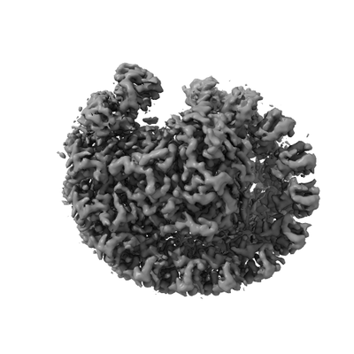 emd_11081.png emd_11081.png | 55.3 KB | ||
| Filedesc metadata | emd-11081.cif.gz | 7.4 KB | ||
| Archive directory |  http://ftp.pdbj.org/pub/emdb/structures/EMD-11081ftp://ftp.pdbj.org/pub/emdb/structures/EMD-11081 http://ftp.pdbj.org/pub/emdb/structures/EMD-11081ftp://ftp.pdbj.org/pub/emdb/structures/EMD-11081 | HTTPS FTP |
-Related structure data
| Related structure data |  6z5sMC  6z5rC M: atomic model generated by this map C: citing same article ( |
|---|---|
| Similar structure data |


-Links
| EMDB pages | EMDB (EBI/PDBe) / EMDataResource |
|---|---|
| Related items in Molecule of the Month |


-Map
| File | Download / File: emd_11081.map.gz / Format: CCP4 / Size: 244.1 MB / Type: IMAGE STORED AS FLOATING POINT NUMBER (4 BYTES) | ||||||||||||||||||||||||||||||||||||||||||||||||||||||||||||
|---|---|---|---|---|---|---|---|---|---|---|---|---|---|---|---|---|---|---|---|---|---|---|---|---|---|---|---|---|---|---|---|---|---|---|---|---|---|---|---|---|---|---|---|---|---|---|---|---|---|---|---|---|---|---|---|---|---|---|---|---|---|
| Projections & slices | Image control
Images are generated by Spider. | ||||||||||||||||||||||||||||||||||||||||||||||||||||||||||||
| Voxel size | X=Y=Z: 1.048 Å | ||||||||||||||||||||||||||||||||||||||||||||||||||||||||||||
| Density |
| ||||||||||||||||||||||||||||||||||||||||||||||||||||||||||||
| Symmetry | Space group: 1 | ||||||||||||||||||||||||||||||||||||||||||||||||||||||||||||
| Details | EMDB XML:
CCP4 map header:
| ||||||||||||||||||||||||||||||||||||||||||||||||||||||||||||
 Z (Sec.)
 Y (Row.)
 X (Col.)


-Supplemental data
- Sample components
+Entire : Reaction center-Light harvesting complex 1-Protein W
+Supramolecule #1: Reaction center-Light harvesting complex 1-Protein W
+Macromolecule #1: Light harvesting complex 1 Protein W
+Macromolecule #2: Reaction center protein M chain
+Macromolecule #3: Reaction center protein L chain
+Macromolecule #4: H subunit of photosynthetic reaction center complex
+Macromolecule #5: Light-harvesting complex 1 alpha chain
+Macromolecule #6: Light-harvesting complex 1 beta chain
+Macromolecule #7: (6~{R},10~{S},14~{R},19~{R},23~{S},24~{E},27~{S},28~{E})-2,6,10,1...

+Macromolecule #8: BACTERIOCHLOROPHYLL A

+Macromolecule #9: BACTERIOPHEOPHYTIN A

+Macromolecule #10: UBIQUINONE-10

+Macromolecule #11: (4S,7R)-4-HYDROXY-N,N,N-TRIMETHYL-9-OXO-7-[(PALMITOYLOXY)METHYL]-...
+Macromolecule #12: CARDIOLIPIN

+Macromolecule #13: FE (III) ION
+Macromolecule #14: DODECYL-BETA-D-MALTOSIDE

+Macromolecule #15: SPIRILLOXANTHIN

+Macromolecule #16: (1S)-2-{[{[(2R)-2,3-DIHYDROXYPROPYL]OXY}(HYDROXY)PHOSPHORYL]OXY}-...
+Macromolecule #17: water

-Experimental details
-Structure determination
| Method | cryo EM |
|---|---|
 Processing Processing | single particle reconstruction |
| Aggregation state | particle |
-Sample preparation
| Buffer | pH: 8 Component:
| ||||||||||||
|---|---|---|---|---|---|---|---|---|---|---|---|---|---|
| Grid | Model: Quantifoil R1.2/1.3 / Material: COPPER / Mesh: 400 / Support film - Material: CARBON / Support film - topology: HOLEY / Pretreatment - Type: GLOW DISCHARGE / Pretreatment - Time: 30 sec. / Pretreatment - Atmosphere: AIR | ||||||||||||
| Vitrification | Cryogen name: ETHANE / Chamber humidity: 60 % / Chamber temperature: 298 K / Instrument: LEICA EM GP | ||||||||||||
| Details | This sample was monodisperse |
- Electron microscopy
| Microscope | FEI TITAN KRIOS |
|---|---|
| Image recording | Film or detector model: GATAN K2 SUMMIT (4k x 4k) / Detector mode: COUNTING / Average electron dose: 46.2 e/Å2 |
| Electron beam | Acceleration voltage: 300 kV / Electron source:  FIELD EMISSION GUN FIELD EMISSION GUN |
| Electron optics | Illumination mode: FLOOD BEAM / Imaging mode: BRIGHT FIELD |
| Experimental equipment |  Model: Titan Krios / Image courtesy: FEI Company |
+Image processing

-Atomic model buiding 1
| Refinement | Space: REAL / Protocol: BACKBONE TRACE |
|---|---|
| Output model | PDB-6z5s: |
