 Movie
Movie Controller

[English] 日本語
 Yorodumi
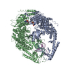
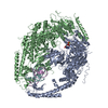
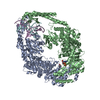
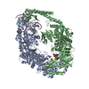
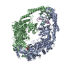
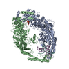
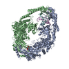
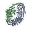
- PDB-2o8e: human MutSalpha (MSH2/MSH6) bound to a G T mispair, with ADP boun... -
+ Open data

- Basic information
| Entry | Database: PDB / ID: 2o8e | ||||||
|---|---|---|---|---|---|---|---|
| Title | human MutSalpha (MSH2/MSH6) bound to a G T mispair, with ADP bound to MSH2 only | ||||||
 Components Components |
| ||||||
 Keywords Keywords | DNA BINDING PROTEIN/DNA / DNA mismatch repair / DNA damage response / somatic hypermutation / protein-DNA complex / DNA mispair / cancer / ABC transporter ATPase / DNA BINDING PROTEIN-DNA COMPLEX | ||||||
| Function / homology |  Function and homology information Function and homology informationsomatic recombination of immunoglobulin genes involved in immune response / MutSbeta complex / Defective Mismatch Repair Associated With MSH3 / MutSalpha complex / Defective Mismatch Repair Associated With MSH6 / Defective Mismatch Repair Associated With MSH2 / somatic recombination of immunoglobulin gene segments / guanine/thymine mispair binding / B cell mediated immunity / maintenance of DNA repeat elements ...somatic recombination of immunoglobulin genes involved in immune response / MutSbeta complex / Defective Mismatch Repair Associated With MSH3 / MutSalpha complex / Defective Mismatch Repair Associated With MSH6 / Defective Mismatch Repair Associated With MSH2 / somatic recombination of immunoglobulin gene segments / guanine/thymine mispair binding / B cell mediated immunity / maintenance of DNA repeat elements / meiotic mismatch repair / histone H3K36me3 reader activity / positive regulation of isotype switching to IgA isotypes / centromeric DNA binding / mismatched DNA binding / positive regulation of isotype switching to IgG isotypes / mitotic recombination / negative regulation of DNA recombination / isotype switching / Mismatch repair (MMR) directed by MSH2:MSH3 (MutSbeta) / Mismatch repair (MMR) directed by MSH2:MSH6 (MutSalpha) / DNA damage tolerance / oxidative phosphorylation / response to UV-B / mitotic intra-S DNA damage checkpoint signaling / ATP-dependent DNA damage sensor activity / germ cell development / intrinsic apoptotic signaling pathway in response to DNA damage by p53 class mediator / response to X-ray / ATP-dependent activity, acting on DNA / somatic hypermutation of immunoglobulin genes / response to UV / mismatch repair / B cell differentiation / intrinsic apoptotic signaling pathway / determination of adult lifespan / TP53 Regulates Transcription of DNA Repair Genes / male gonad development / intrinsic apoptotic signaling pathway in response to DNA damage / enzyme activator activity / double-strand break repair / in utero embryonic development / spermatogenesis / negative regulation of neuron apoptotic process / damaged DNA binding / chromosome, telomeric region / DNA repair / chromatin binding / chromatin / Golgi apparatus / enzyme binding / protein homodimerization activity / ATP hydrolysis activity / DNA binding / nucleoplasm / ATP binding / membrane / nucleus / cytosol Similarity search - Function | ||||||
| Biological species |  Homo sapiens (human) Homo sapiens (human) | ||||||
| Method |  X-RAY DIFFRACTION / SYNCHROTRON / FOURIER SYNTHESIS / Resolution: 3.3 Å X-RAY DIFFRACTION / SYNCHROTRON / FOURIER SYNTHESIS / Resolution: 3.3 Å | ||||||
 Authors Authors | Warren, J.J. / Pohlhaus, T.J. / Changela, A. / Modrich, P.L. / Beese, L.S. | ||||||
 Citation Citation | Journal: Mol.Cell / Year: 2007 Title: Structure of the Human MutSalpha DNA Lesion Recognition Complex. Authors: Warren, J.J. / Pohlhaus, T.J. / Changela, A. / Iyer, R.R. / Modrich, P.L. / Beese, L.S. | ||||||
| History |
|
- Structure visualization
| Structure viewer | Molecule: MolmilJmol/JSmol |
|---|
- Downloads & links
-Download
| PDBx/mmCIF format | 2o8e.cif.gz | 377.4 KB | Display | PDBx/mmCIF format |
|---|---|---|---|---|
| PDB format | pdb2o8e.ent.gz | 291.9 KB | Display | PDB format |
| PDBx/mmJSON format | 2o8e.json.gz | Tree view | PDBx/mmJSON format | |
| Others |  Other downloads Other downloads |
-Validation report
| Arichive directory | https://data.pdbj.org/pub/pdb/validation_reports/o8/2o8eftp://data.pdbj.org/pub/pdb/validation_reports/o8/2o8e | HTTPS FTP |
|---|
-Related structure data
| Related structure data |  2o8bSC 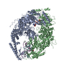 2o8cC  2o8dC  2o8fC S: Starting model for refinement C: citing same article ( |
|---|---|
| Similar structure data |


-Links
 PDBj
PDBj


- Assembly
| Deposited unit | 
| ||||||||
|---|---|---|---|---|---|---|---|---|---|
| 1 |
| ||||||||
| Unit cell |
| ||||||||
| Components on special symmetry positions |
| ||||||||
| Details | the crystallographic asymmetric unit contains one biological assembly (a heterodimeric protein complex bound to double stranded DNA and one ADP+Mg) |
-Components
-DNA chain , 2 types, 2 molecules EF
| #1: DNA chain | Mass: 4660.023 Da / Num. of mol.: 1 / Mutation: F42A / Source method: obtained synthetically |
|---|---|
| #2: DNA chain | Mass: 4536.934 Da / Num. of mol.: 1 / Source method: obtained synthetically |
-DNA mismatch repair protein ... , 2 types, 2 molecules AB
| #3: Protein | Mass: 104785.781 Da / Num. of mol.: 1 Source method: isolated from a genetically manipulated source Source: (gene. exp.) Homo sapiens (human) / Gene: MSH2 / Plasmid: pFasBacDual / Production host:   Spodoptera frugiperda (fall armyworm) / Strain (production host): SF9 / References: UniProt: P43246 Spodoptera frugiperda (fall armyworm) / Strain (production host): SF9 / References: UniProt: P43246 |
|---|---|
| #4: Protein | Mass: 116056.672 Da / Num. of mol.: 1 / Fragment: residues 341-1360 Source method: isolated from a genetically manipulated source Source: (gene. exp.) Homo sapiens (human) / Gene: MSH6, GTBP / Plasmid: pFasBacDual / Production host: Spodoptera frugiperda (fall armyworm) / Strain (production host): sf9 / References: UniProt: P52701 |
-Non-polymers , 3 types, 12 molecules 


| #5: Chemical | ChemComp-MG /  Mass: 24.305 Da / Num. of mol.: 1 / Source method: obtained synthetically / Formula: Mg Mass: 24.305 Da / Num. of mol.: 1 / Source method: obtained synthetically / Formula: Mg |
|---|---|
| #6: Chemical | ChemComp-ADP /  Mass: 427.201 Da / Num. of mol.: 1 / Source method: obtained synthetically / Formula: C10H15N5O10P2 / Comment: ADP, energy-carrying molecule*YM Mass: 427.201 Da / Num. of mol.: 1 / Source method: obtained synthetically / Formula: C10H15N5O10P2 / Comment: ADP, energy-carrying molecule*YM |
| #7: Water | ChemComp-HOH / Mass: 18.015 Da / Num. of mol.: 10 / Source method: isolated from a natural source / Formula: H2O |
-Experimental details
-Experiment
| Experiment | Method: X-RAY DIFFRACTION / Number of used crystals: 1 |
|---|
- Sample preparation
| Crystal | Density Matthews: 3.09 Å3/Da / Density % sol: 60.15 % | ||||||||||||||||||||||||||||||||||||
|---|---|---|---|---|---|---|---|---|---|---|---|---|---|---|---|---|---|---|---|---|---|---|---|---|---|---|---|---|---|---|---|---|---|---|---|---|---|
| Crystal grow | Temperature: 290 K / Method: vapor diffusion / pH: 7 Details: 11% PEG 8000, 10 mM magnesium sulfate, 100 mM bis-tris-propane, pH 7, VAPOR DIFFUSION, temperature 290K | ||||||||||||||||||||||||||||||||||||
| Components of the solutions |
|
-Data collection
| Diffraction | Mean temperature: 100 K | ||||||||||||||||||||||||||||||||||||||||||||||||||||||||||||||||||||||||||||||||||||||||||||||||||||||||||||||||||||||||||||||||||||||||||||||||||||||||||||||||||||||||
|---|---|---|---|---|---|---|---|---|---|---|---|---|---|---|---|---|---|---|---|---|---|---|---|---|---|---|---|---|---|---|---|---|---|---|---|---|---|---|---|---|---|---|---|---|---|---|---|---|---|---|---|---|---|---|---|---|---|---|---|---|---|---|---|---|---|---|---|---|---|---|---|---|---|---|---|---|---|---|---|---|---|---|---|---|---|---|---|---|---|---|---|---|---|---|---|---|---|---|---|---|---|---|---|---|---|---|---|---|---|---|---|---|---|---|---|---|---|---|---|---|---|---|---|---|---|---|---|---|---|---|---|---|---|---|---|---|---|---|---|---|---|---|---|---|---|---|---|---|---|---|---|---|---|---|---|---|---|---|---|---|---|---|---|---|---|---|---|---|---|
| Diffraction source | Source: SYNCHROTRON / Site: APS  / Beamline: 22-ID / Wavelength: 1.005 Å / Beamline: 22-ID / Wavelength: 1.005 Å | ||||||||||||||||||||||||||||||||||||||||||||||||||||||||||||||||||||||||||||||||||||||||||||||||||||||||||||||||||||||||||||||||||||||||||||||||||||||||||||||||||||||||
| Detector | Type: MARMOSAIC 300 mm CCD / Detector: CCD / Details: mirrors | ||||||||||||||||||||||||||||||||||||||||||||||||||||||||||||||||||||||||||||||||||||||||||||||||||||||||||||||||||||||||||||||||||||||||||||||||||||||||||||||||||||||||
| Radiation | Monochromator: Si 220 / Protocol: SINGLE WAVELENGTH / Monochromatic (M) / Laue (L): M / Scattering type: x-ray | ||||||||||||||||||||||||||||||||||||||||||||||||||||||||||||||||||||||||||||||||||||||||||||||||||||||||||||||||||||||||||||||||||||||||||||||||||||||||||||||||||||||||
| Radiation wavelength | Wavelength: 1.005 Å / Relative weight: 1 | ||||||||||||||||||||||||||||||||||||||||||||||||||||||||||||||||||||||||||||||||||||||||||||||||||||||||||||||||||||||||||||||||||||||||||||||||||||||||||||||||||||||||
| Reflection | Resolution: 3.3→91.044 Å / Num. all: 43653 / Num. obs: 43653 / % possible obs: 98.9 % / Observed criterion σ(I): -3 / Redundancy: 9.1 % / Rmerge(I) obs: 0.096 / Rsym value: 0.096 / Net I/σ(I): 6.6 | ||||||||||||||||||||||||||||||||||||||||||||||||||||||||||||||||||||||||||||||||||||||||||||||||||||||||||||||||||||||||||||||||||||||||||||||||||||||||||||||||||||||||
| Reflection shell | Diffraction-ID: 1
|
- Processing
| Software |
| ||||||||||||||||||||||||||||||||||||||||||||||||||||||||||||||||||||||||||||||||||||||||||||||||||||||||||||||||||||||||||||||||||||||||||||||||||||||||||||||||||||||||||||||||||||||||||||||||||||||||||||||||||||||||||||||||||||||||||||||||||||||||||||||||||||||||||||||||||||||||||||||||||||||||||||
|---|---|---|---|---|---|---|---|---|---|---|---|---|---|---|---|---|---|---|---|---|---|---|---|---|---|---|---|---|---|---|---|---|---|---|---|---|---|---|---|---|---|---|---|---|---|---|---|---|---|---|---|---|---|---|---|---|---|---|---|---|---|---|---|---|---|---|---|---|---|---|---|---|---|---|---|---|---|---|---|---|---|---|---|---|---|---|---|---|---|---|---|---|---|---|---|---|---|---|---|---|---|---|---|---|---|---|---|---|---|---|---|---|---|---|---|---|---|---|---|---|---|---|---|---|---|---|---|---|---|---|---|---|---|---|---|---|---|---|---|---|---|---|---|---|---|---|---|---|---|---|---|---|---|---|---|---|---|---|---|---|---|---|---|---|---|---|---|---|---|---|---|---|---|---|---|---|---|---|---|---|---|---|---|---|---|---|---|---|---|---|---|---|---|---|---|---|---|---|---|---|---|---|---|---|---|---|---|---|---|---|---|---|---|---|---|---|---|---|---|---|---|---|---|---|---|---|---|---|---|---|---|---|---|---|---|---|---|---|---|---|---|---|---|---|---|---|---|---|---|---|---|---|---|---|---|---|---|---|---|---|---|---|---|---|---|---|---|---|---|---|---|---|---|---|---|---|---|---|---|---|---|---|---|---|---|---|---|---|---|---|---|---|---|---|---|---|---|---|---|---|---|
| Refinement | Method to determine structure: FOURIER SYNTHESIS Starting model: PDB ENTRY 2O8B Resolution: 3.3→20 Å / Cor.coef. Fo:Fc: 0.89 / Cor.coef. Fo:Fc free: 0.858 / SU B: 62.226 / SU ML: 0.49 / TLS residual ADP flag: LIKELY RESIDUAL / Isotropic thermal model: TLS refinement by domains / Cross valid method: THROUGHOUT / σ(F): 0 / ESU R Free: 0.584 / Stereochemistry target values: MAXIMUM LIKELIHOOD / Details: HYDROGENS HAVE BEEN ADDED IN THE RIDING POSITIONS
| ||||||||||||||||||||||||||||||||||||||||||||||||||||||||||||||||||||||||||||||||||||||||||||||||||||||||||||||||||||||||||||||||||||||||||||||||||||||||||||||||||||||||||||||||||||||||||||||||||||||||||||||||||||||||||||||||||||||||||||||||||||||||||||||||||||||||||||||||||||||||||||||||||||||||||||
| Solvent computation | Ion probe radii: 0.8 Å / Shrinkage radii: 0.8 Å / VDW probe radii: 1.2 Å / Solvent model: MASK | ||||||||||||||||||||||||||||||||||||||||||||||||||||||||||||||||||||||||||||||||||||||||||||||||||||||||||||||||||||||||||||||||||||||||||||||||||||||||||||||||||||||||||||||||||||||||||||||||||||||||||||||||||||||||||||||||||||||||||||||||||||||||||||||||||||||||||||||||||||||||||||||||||||||||||||
| Displacement parameters | Biso mean: 103.516 Å2 | ||||||||||||||||||||||||||||||||||||||||||||||||||||||||||||||||||||||||||||||||||||||||||||||||||||||||||||||||||||||||||||||||||||||||||||||||||||||||||||||||||||||||||||||||||||||||||||||||||||||||||||||||||||||||||||||||||||||||||||||||||||||||||||||||||||||||||||||||||||||||||||||||||||||||||||
| Refinement step | Cycle: LAST / Resolution: 3.3→20 Å
| ||||||||||||||||||||||||||||||||||||||||||||||||||||||||||||||||||||||||||||||||||||||||||||||||||||||||||||||||||||||||||||||||||||||||||||||||||||||||||||||||||||||||||||||||||||||||||||||||||||||||||||||||||||||||||||||||||||||||||||||||||||||||||||||||||||||||||||||||||||||||||||||||||||||||||||
| Refine LS restraints |
| ||||||||||||||||||||||||||||||||||||||||||||||||||||||||||||||||||||||||||||||||||||||||||||||||||||||||||||||||||||||||||||||||||||||||||||||||||||||||||||||||||||||||||||||||||||||||||||||||||||||||||||||||||||||||||||||||||||||||||||||||||||||||||||||||||||||||||||||||||||||||||||||||||||||||||||
| LS refinement shell | Resolution: 3.3→3.383 Å / Total num. of bins used: 20
| ||||||||||||||||||||||||||||||||||||||||||||||||||||||||||||||||||||||||||||||||||||||||||||||||||||||||||||||||||||||||||||||||||||||||||||||||||||||||||||||||||||||||||||||||||||||||||||||||||||||||||||||||||||||||||||||||||||||||||||||||||||||||||||||||||||||||||||||||||||||||||||||||||||||||||||
| Refinement TLS params. | Method: refined / Refine-ID: X-RAY DIFFRACTION
| ||||||||||||||||||||||||||||||||||||||||||||||||||||||||||||||||||||||||||||||||||||||||||||||||||||||||||||||||||||||||||||||||||||||||||||||||||||||||||||||||||||||||||||||||||||||||||||||||||||||||||||||||||||||||||||||||||||||||||||||||||||||||||||||||||||||||||||||||||||||||||||||||||||||||||||
| Refinement TLS group |
|
