 Movie
Movie Controller

[English] 日本語
 Yorodumi
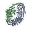
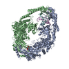
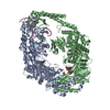
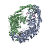
- PDB-2wtu: Crystal structure of Escherichia coli MutS in complex with a 16 b... -
+ Open data

- Basic information
| Entry | Database: PDB / ID: 2wtu | ||||||
|---|---|---|---|---|---|---|---|
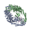
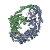
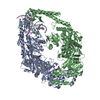
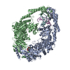
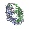
| Title | Crystal structure of Escherichia coli MutS in complex with a 16 basepair oligo containing an A.A mismatch. | ||||||
 Components Components |
| ||||||
 Keywords Keywords | DNA BINDING PROTEIN/DNA / DNA BINDING PROTEIN-DNA COMPLEX / DNA DAMAGE / DNA REPAIR / NUCLEOTIDE-BINDING | ||||||
| Function / homology |  Function and homology information Function and homology informationadenine/cytosine mispair binding / MutS complex / mismatch repair complex / regulation of DNA recombination / mismatched DNA binding / DNA binding, bending / ATP-dependent DNA damage sensor activity / mismatch repair / ADP binding / damaged DNA binding ...adenine/cytosine mispair binding / MutS complex / mismatch repair complex / regulation of DNA recombination / mismatched DNA binding / DNA binding, bending / ATP-dependent DNA damage sensor activity / mismatch repair / ADP binding / damaged DNA binding / DNA damage response / ATP hydrolysis activity / ATP binding / identical protein binding / cytosol Similarity search - Function | ||||||
| Biological species |  synthetic construct (others) | ||||||
| Method |  X-RAY DIFFRACTION / SYNCHROTRON / MOLECULAR REPLACEMENT / Resolution: 3.4 Å X-RAY DIFFRACTION / SYNCHROTRON / MOLECULAR REPLACEMENT / Resolution: 3.4 Å | ||||||
 Authors Authors | Natrajan, G. / Lebbink, J.H. / Reumer, A. / Fish, A. / Winterwerp, H.H. / Sixma, T.K. | ||||||
 Citation Citation | Journal: J. Biol. Chem. / Year: 2010 Title: Magnesium coordination controls the molecular switch function of DNA mismatch repair protein MutS. Authors: Lebbink, J.H. / Fish, A. / Reumer, A. / Natrajan, G. / Winterwerp, H.H. / Sixma, T.K. | ||||||
| History |
|
- Structure visualization
| Structure viewer | Molecule: MolmilJmol/JSmol |
|---|
- Downloads & links
-Download
| PDBx/mmCIF format | 2wtu.cif.gz | 320.9 KB | Display | PDBx/mmCIF format |
|---|---|---|---|---|
| PDB format | pdb2wtu.ent.gz | 254.9 KB | Display | PDB format |
| PDBx/mmJSON format | 2wtu.json.gz | Tree view | PDBx/mmJSON format | |
| Others |  Other downloads Other downloads |
-Validation report
| Arichive directory | https://data.pdbj.org/pub/pdb/validation_reports/wt/2wtuftp://data.pdbj.org/pub/pdb/validation_reports/wt/2wtu | HTTPS FTP |
|---|
-Related structure data
| Related structure data |  3k0sC  1oh6S S: Starting model for refinement C: citing same article ( |
|---|---|
| Similar structure data |


-Links
 PDBj
PDBj


- Assembly
| Deposited unit | 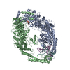
| ||||||||
|---|---|---|---|---|---|---|---|---|---|
| 1 |
| ||||||||
| Unit cell |
|
-Components
-Protein , 1 types, 2 molecules AB
| #1: Protein | Mass: 89604.359 Da / Num. of mol.: 2 / Fragment: RESIDUES 1-800 Source method: isolated from a genetically manipulated source Source: (gene. exp.) |
|---|
-DNA chain , 2 types, 2 molecules EF
| #2: DNA chain | Mass: 4877.183 Da / Num. of mol.: 1 / Source method: obtained synthetically / Details: THIS IS A CHEMICALLY SYNTHESIZED OLIGONUCLEOTIDE. / Source: (synth.) synthetic construct (others) |
|---|---|
| #3: DNA chain | Mass: 4930.189 Da / Num. of mol.: 1 / Source method: obtained synthetically / Details: THIS IS A CHEMICALLY SYNTHESIZED OLIGONUCLEOTIDE. / Source: (synth.) synthetic construct (others) |
-Non-polymers , 3 types, 45 molecules 


| #4: Chemical | ChemComp-ADP /  Mass: 427.201 Da / Num. of mol.: 1 / Source method: obtained synthetically / Formula: C10H15N5O10P2 / Comment: ADP, energy-carrying molecule*YM Mass: 427.201 Da / Num. of mol.: 1 / Source method: obtained synthetically / Formula: C10H15N5O10P2 / Comment: ADP, energy-carrying molecule*YM | ||
|---|---|---|---|
| #5: Chemical |  Mass: 35.453 Da / Num. of mol.: 3 / Source method: obtained synthetically / Formula: Cl Mass: 35.453 Da / Num. of mol.: 3 / Source method: obtained synthetically / Formula: Cl#6: Water | ChemComp-HOH / | Mass: 18.015 Da / Num. of mol.: 41 / Source method: isolated from a natural source / Formula: H2O |
-Details
| Sequence details | THE CRYSTALLIZ |
|---|
-Experimental details
-Experiment
| Experiment | Method: X-RAY DIFFRACTION / Number of used crystals: 1 |
|---|
- Sample preparation
| Crystal | Density Matthews: 2.82 Å3/Da / Density % sol: 56.36 % / Description: NONE |
|---|---|
| Crystal grow | pH: 7.5 Details: 18% PEG 3350, 100 MM SODIUM CITRATE, 100 MM BIS TRIS PROPANE PH 7.5, 5 MM MGCL2, 100 MICROM ADP. |
-Data collection
| Diffraction | Mean temperature: 100 K |
|---|---|
| Diffraction source | Source: SYNCHROTRON / Site: ESRF  / Beamline: ID14-3 / Wavelength: 0.93 / Beamline: ID14-3 / Wavelength: 0.93 |
| Detector | Type: ADSC CCD / Detector: CCD / Date: Jun 15, 2004 / Details: TOROIDAL MIRRORS |
| Radiation | Monochromator: SI 111 / Protocol: SINGLE WAVELENGTH / Monochromatic (M) / Laue (L): M / Scattering type: x-ray |
| Radiation wavelength | Wavelength: 0.93 Å / Relative weight: 1 |
| Reflection | Resolution: 3.4→50 Å / Num. obs: 28641 / % possible obs: 99.8 % / Observed criterion σ(I): 1.1 / Redundancy: 7.2 % / Biso Wilson estimate: 72.1 Å2 / Rmerge(I) obs: 0.15 / Net I/σ(I): 13.6 |
| Reflection shell | Resolution: 3.4→3.58 Å / Redundancy: 7.2 % / Rmerge(I) obs: 0.47 / Mean I/σ(I) obs: 4.3 / % possible all: 99.8 |
- Processing
| Software |
| ||||||||||||||||||||||||||||||||||||||||||||||||||||||||||||||||||||||||||||||||||||||||||||||||||||||||||||||||||||||||||||||||||||||||||||||||||||||||||||||||||||||||||||||||||||||
|---|---|---|---|---|---|---|---|---|---|---|---|---|---|---|---|---|---|---|---|---|---|---|---|---|---|---|---|---|---|---|---|---|---|---|---|---|---|---|---|---|---|---|---|---|---|---|---|---|---|---|---|---|---|---|---|---|---|---|---|---|---|---|---|---|---|---|---|---|---|---|---|---|---|---|---|---|---|---|---|---|---|---|---|---|---|---|---|---|---|---|---|---|---|---|---|---|---|---|---|---|---|---|---|---|---|---|---|---|---|---|---|---|---|---|---|---|---|---|---|---|---|---|---|---|---|---|---|---|---|---|---|---|---|---|---|---|---|---|---|---|---|---|---|---|---|---|---|---|---|---|---|---|---|---|---|---|---|---|---|---|---|---|---|---|---|---|---|---|---|---|---|---|---|---|---|---|---|---|---|---|---|---|---|
| Refinement | Method to determine structure: MOLECULAR REPLACEMENT Starting model: PDB ENTRY 1OH6 Resolution: 3.4→50 Å / Cor.coef. Fo:Fc: 0.923 / Cor.coef. Fo:Fc free: 0.88 / SU B: 30.691 / SU ML: 0.491 / Cross valid method: THROUGHOUT / ESU R Free: 0.624 / Stereochemistry target values: MAXIMUM LIKELIHOOD Details: HYDROGENS HAVE BEEN ADDED IN THE RIDING POSITIONS. U VALUES REFINED INDIVIDUALLY. MISSING RESIDUES ARE DUE TO LACK OF VISIBLE ELECTRON DENSITY.
| ||||||||||||||||||||||||||||||||||||||||||||||||||||||||||||||||||||||||||||||||||||||||||||||||||||||||||||||||||||||||||||||||||||||||||||||||||||||||||||||||||||||||||||||||||||||
| Solvent computation | Ion probe radii: 0.8 Å / Shrinkage radii: 0.8 Å / VDW probe radii: 1.4 Å / Solvent model: MASK | ||||||||||||||||||||||||||||||||||||||||||||||||||||||||||||||||||||||||||||||||||||||||||||||||||||||||||||||||||||||||||||||||||||||||||||||||||||||||||||||||||||||||||||||||||||||
| Displacement parameters | Biso mean: 52.791 Å2
| ||||||||||||||||||||||||||||||||||||||||||||||||||||||||||||||||||||||||||||||||||||||||||||||||||||||||||||||||||||||||||||||||||||||||||||||||||||||||||||||||||||||||||||||||||||||
| Refinement step | Cycle: LAST / Resolution: 3.4→50 Å
| ||||||||||||||||||||||||||||||||||||||||||||||||||||||||||||||||||||||||||||||||||||||||||||||||||||||||||||||||||||||||||||||||||||||||||||||||||||||||||||||||||||||||||||||||||||||
| Refine LS restraints |
|