 Movie
Movie Controller

[English] 日本語
 Yorodumi
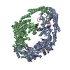
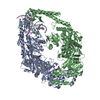
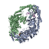
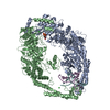
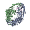
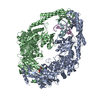
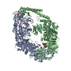
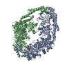
- PDB-3zlj: CRYSTAL STRUCTURE OF FULL-LENGTH E.COLI DNA MISMATCH REPAIR PROTE... -
+ Open data

- Basic information
| Entry | Database: PDB / ID: 3zlj | ||||||
|---|---|---|---|---|---|---|---|
| Title | CRYSTAL STRUCTURE OF FULL-LENGTH E.COLI DNA MISMATCH REPAIR PROTEIN MUTS D835R MUTANT IN COMPLEX WITH GT MISMATCHED DNA | ||||||
 Components Components |
| ||||||
 Keywords Keywords | DNA BINDING PROTEIN/DNA / DNA BINDING PROTEIN-DNA COMPLEX / DIMER MUTANT / MISMATCH REPAIR / DNA REPAIR PROTEIN / DNA DAMAGE / NUCLEOTIDE-BINDING / ATP-BINDING | ||||||
| Function / homology |  Function and homology information Function and homology informationadenine/cytosine mispair binding / MutS complex / mismatch repair complex / regulation of DNA recombination / mismatched DNA binding / DNA binding, bending / ATP-dependent DNA damage sensor activity / mismatch repair / ADP binding / damaged DNA binding ...adenine/cytosine mispair binding / MutS complex / mismatch repair complex / regulation of DNA recombination / mismatched DNA binding / DNA binding, bending / ATP-dependent DNA damage sensor activity / mismatch repair / ADP binding / damaged DNA binding / DNA damage response / ATP hydrolysis activity / ATP binding / identical protein binding / cytosol Similarity search - Function | ||||||
| Biological species |  SYNTHETIC CONSTRUCT (others) | ||||||
| Method |  X-RAY DIFFRACTION / SYNCHROTRON / MOLECULAR REPLACEMENT / Resolution: 3.1 Å X-RAY DIFFRACTION / SYNCHROTRON / MOLECULAR REPLACEMENT / Resolution: 3.1 Å | ||||||
 Authors Authors | Groothuizen, F.S. / Fish, A. / Petoukhov, M.V. / Reumer, A. / Manelyte, L. / Winterwerp, H.H.K. / Marinus, M.G. / Lebbink, J.H.G. / Svergun, D.I. / Friedhoff, P. / Sixma, T.K. | ||||||
 Citation Citation | Journal: Nucleic Acids Res / Year: 2013 Title: Using stable MutS dimers and tetramers to quantitatively analyze DNA mismatch recognition and sliding clamp formation. Authors: Flora S Groothuizen / Alexander Fish / Maxim V Petoukhov / Annet Reumer / Laura Manelyte / Herrie H K Winterwerp / Martin G Marinus / Joyce H G Lebbink / Dmitri I Svergun / Peter Friedhoff / Titia K Sixma /  Abstract: The process of DNA mismatch repair is initiated when MutS recognizes mismatched DNA bases and starts the repair cascade. The Escherichia coli MutS protein exists in an equilibrium between dimers and ...The process of DNA mismatch repair is initiated when MutS recognizes mismatched DNA bases and starts the repair cascade. The Escherichia coli MutS protein exists in an equilibrium between dimers and tetramers, which has compromised biophysical analysis. To uncouple these states, we have generated stable dimers and tetramers, respectively. These proteins allowed kinetic analysis of DNA recognition and structural analysis of the full-length protein by X-ray crystallography and small angle X-ray scattering. Our structural data reveal that the tetramerization domains are flexible with respect to the body of the protein, resulting in mostly extended structures. Tetrameric MutS has a slow dissociation from DNA, which can be due to occasional bending over and binding DNA in its two binding sites. In contrast, the dimer dissociation is faster, primarily dependent on a combination of the type of mismatch and the flanking sequence. In the presence of ATP, we could distinguish two kinetic groups: DNA sequences where MutS forms sliding clamps and those where sliding clamps are not formed efficiently. Interestingly, this inability to undergo a conformational change rather than mismatch affinity is correlated with mismatch repair. | ||||||
| History |
|
- Structure visualization
| Structure viewer | Molecule: MolmilJmol/JSmol |
|---|
- Downloads & links
-Download
| PDBx/mmCIF format | 3zlj.cif.gz | 666.7 KB | Display | PDBx/mmCIF format |
|---|---|---|---|---|
| PDB format | pdb3zlj.ent.gz | 551.7 KB | Display | PDB format |
| PDBx/mmJSON format | 3zlj.json.gz | Tree view | PDBx/mmJSON format | |
| Others |  Other downloads Other downloads |
-Validation report
| Arichive directory | https://data.pdbj.org/pub/pdb/validation_reports/zl/3zljftp://data.pdbj.org/pub/pdb/validation_reports/zl/3zlj | HTTPS FTP |
|---|
-Related structure data
| Related structure data |  1e3mS S: Starting model for refinement C: citing same article ( |
|---|---|
| Similar structure data |


-Links
 PDBj
PDBj


- Assembly
| Deposited unit | 
| ||||||||||||||||||||||||||||||||||||||||||||||||||||||||||||||||||||
|---|---|---|---|---|---|---|---|---|---|---|---|---|---|---|---|---|---|---|---|---|---|---|---|---|---|---|---|---|---|---|---|---|---|---|---|---|---|---|---|---|---|---|---|---|---|---|---|---|---|---|---|---|---|---|---|---|---|---|---|---|---|---|---|---|---|---|---|---|---|
| 1 | 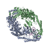
| ||||||||||||||||||||||||||||||||||||||||||||||||||||||||||||||||||||
| 2 | 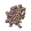
| ||||||||||||||||||||||||||||||||||||||||||||||||||||||||||||||||||||
| Unit cell |
| ||||||||||||||||||||||||||||||||||||||||||||||||||||||||||||||||||||
| Noncrystallographic symmetry (NCS) | NCS domain:
NCS domain segments: Component-ID: _ / Refine code: _
NCS ensembles :
|
-Components
| #1: Protein | Mass: 89604.359 Da / Num. of mol.: 2 / Mutation: YES Source method: isolated from a genetically manipulated source Source: (gene. exp.) #2: Protein | Mass: 5811.530 Da / Num. of mol.: 2 / Mutation: YES Source method: isolated from a genetically manipulated source Details: CHAINS C AND D ARE THE C-TERMINAL PORTION OF CHAINS A AND B, HOWEVER THE MISSING REGION 800-822 MAKES UNAMBIGUOUS ASSIGNMENT TO THE CORRECT CHAIN IMPOSSIBLE Source: (gene. exp.) #3: DNA chain | | Mass: 6433.162 Da / Num. of mol.: 1 / Source method: obtained synthetically / Source: (synth.) SYNTHETIC CONSTRUCT (others) #4: DNA chain | | Mass: 6470.169 Da / Num. of mol.: 1 / Source method: obtained synthetically / Source: (synth.) SYNTHETIC CONSTRUCT (others) |
|---|
-Experimental details
-Experiment
| Experiment | Method: X-RAY DIFFRACTION / Number of used crystals: 1 |
|---|
- Sample preparation
| Crystal | Density Matthews: 2.72 Å3/Da / Density % sol: 59.7 % / Description: NONE |
|---|---|
| Crystal grow | Temperature: 293 K / Method: vapor diffusion, hanging drop / pH: 7.5 Details: 5MM TRIS PH8, 750MM NACL, 12% PEG 6000, 10MM MGCL2, VAPOR DIFFUSION, HANGING DROP, TEMPERATURE 293K, pH 7.5 |
-Data collection
| Diffraction | Mean temperature: 100 K |
|---|---|
| Diffraction source | Source: SYNCHROTRON / Site: ESRF  / Beamline: ID14-4 / Wavelength: 0.976 / Beamline: ID14-4 / Wavelength: 0.976 |
| Detector | Type: ADSC QUANTUM 315r / Detector: CCD / Date: May 2, 2008 |
| Radiation | Protocol: SINGLE WAVELENGTH / Monochromatic (M) / Laue (L): M / Scattering type: x-ray |
| Radiation wavelength | Wavelength: 0.976 Å / Relative weight: 1 |
| Reflection | Resolution: 3.1→47.24 Å / Num. obs: 40031 / % possible obs: 99.9 % / Observed criterion σ(I): 2 / Redundancy: 4.4 % / Rmerge(I) obs: 0.12 / Net I/σ(I): 9.9 |
| Reflection shell | Resolution: 3.1→3.27 Å / Redundancy: 4.4 % / Rmerge(I) obs: 0.62 / Mean I/σ(I) obs: 2.4 / % possible all: 100 |
- Processing
| Software |
| ||||||||||||||||||||||||||||||||||||||||||||||||||||||||||||||||||||||||||||||||||||||||||||||||||||||||||||||||||||||||||||||||||||||||||||||||||||||||||||||||||||||||||||||||||||||
|---|---|---|---|---|---|---|---|---|---|---|---|---|---|---|---|---|---|---|---|---|---|---|---|---|---|---|---|---|---|---|---|---|---|---|---|---|---|---|---|---|---|---|---|---|---|---|---|---|---|---|---|---|---|---|---|---|---|---|---|---|---|---|---|---|---|---|---|---|---|---|---|---|---|---|---|---|---|---|---|---|---|---|---|---|---|---|---|---|---|---|---|---|---|---|---|---|---|---|---|---|---|---|---|---|---|---|---|---|---|---|---|---|---|---|---|---|---|---|---|---|---|---|---|---|---|---|---|---|---|---|---|---|---|---|---|---|---|---|---|---|---|---|---|---|---|---|---|---|---|---|---|---|---|---|---|---|---|---|---|---|---|---|---|---|---|---|---|---|---|---|---|---|---|---|---|---|---|---|---|---|---|---|---|
| Refinement | Method to determine structure: MOLECULAR REPLACEMENT Starting model: PDB ENTRY 1E3M Resolution: 3.1→46.49 Å / Cor.coef. Fo:Fc: 0.917 / Cor.coef. Fo:Fc free: 0.884 / SU B: 49.479 / SU ML: 0.38 / Cross valid method: THROUGHOUT / ESU R Free: 0.49 / Stereochemistry target values: MAXIMUM LIKELIHOOD Details: HYDROGENS HAVE BEEN ADDED IN THE RIDING POSITIONS. U VALUES WITH TLS ADDED
| ||||||||||||||||||||||||||||||||||||||||||||||||||||||||||||||||||||||||||||||||||||||||||||||||||||||||||||||||||||||||||||||||||||||||||||||||||||||||||||||||||||||||||||||||||||||
| Solvent computation | Ion probe radii: 0.9 Å / Shrinkage radii: 0.9 Å / VDW probe radii: 1 Å / Solvent model: MASK | ||||||||||||||||||||||||||||||||||||||||||||||||||||||||||||||||||||||||||||||||||||||||||||||||||||||||||||||||||||||||||||||||||||||||||||||||||||||||||||||||||||||||||||||||||||||
| Displacement parameters | Biso mean: 61.792 Å2
| ||||||||||||||||||||||||||||||||||||||||||||||||||||||||||||||||||||||||||||||||||||||||||||||||||||||||||||||||||||||||||||||||||||||||||||||||||||||||||||||||||||||||||||||||||||||
| Refinement step | Cycle: LAST / Resolution: 3.1→46.49 Å
| ||||||||||||||||||||||||||||||||||||||||||||||||||||||||||||||||||||||||||||||||||||||||||||||||||||||||||||||||||||||||||||||||||||||||||||||||||||||||||||||||||||||||||||||||||||||
| Refine LS restraints |
|