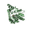
Arachidonate production from DAG / Acyl chain remodeling of DAG and TAG / acylglycerol catabolic process / acylglycerol lipase / monoacylglycerol catabolic process / regulation of endocannabinoid signaling pathway / monoacylglycerol lipase activity / arachidonate metabolic process / regulation of sensory perception of pain / phosphatidylcholine lysophospholipase A1 activity ...Arachidonate production from DAG / Acyl chain remodeling of DAG and TAG / acylglycerol catabolic process / acylglycerol lipase / monoacylglycerol catabolic process / regulation of endocannabinoid signaling pathway / monoacylglycerol lipase activity / arachidonate metabolic process / regulation of sensory perception of pain / phosphatidylcholine lysophospholipase A1 activity / triglyceride catabolic process / Triglyceride catabolism / regulation of signal transduction / lipid metabolic process / fatty acid biosynthetic process / MLL4 and MLL3 complexes regulate expression of PPARG target genes in adipogenesis and hepatic steatosis / regulation of inflammatory response / inflammatory response / endoplasmic reticulum membrane / protein homodimerization activity / membrane / plasma membrane / cytosol Similarity search - Function
Mass: 18.015 Da / Num. of mol.: 409 / Source method: isolated from a natural source / Formula: H2O
Has protein modification
Y
-
Experimental details
-
Experiment
Experiment
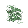
Method: X-RAY DIFFRACTION / Number of used crystals: 1
-
Sample preparation
Crystal
Density Matthews: 2.46 Å3/Da / Density % sol: 50.04 %
Crystal grow
Temperature: 298 K / Method: vapor diffusion, sitting drop Details: Reservoir Buffer: 0.07M sodium cacodylate pH 5.1-5.9 and 33-51% MPD The protein is 20 mg/mL in a buffer of 15 mM HEPES pH 8.2; 2 mM TCEP; and 10% glycerol, with hexaethylene glycol ...Details: Reservoir Buffer: 0.07M sodium cacodylate pH 5.1-5.9 and 33-51% MPD The protein is 20 mg/mL in a buffer of 15 mM HEPES pH 8.2; 2 mM TCEP; and 10% glycerol, with hexaethylene glycol monododecyl ether added to 0.1 mM. Apo protein crystals obtained in this manner were transferred to a cryo-protectant solution consisting of 70 mM NaCacodylate pH 5.1; 10% MPD; 30% PEG-MME-2K, and 1mM of inhibitor compound. Crystals were soaked overnight, then flash-frozen in LN2. PH range: 5.1 - 5.9
In the structure databanks used in Yorodumi, some data are registered as the other names, "COVID-19 virus" and "2019-nCoV". Here are the details of the virus and the list of structure data.
Jan 31, 2019. EMDB accession codes are about to change! (news from PDBe EMDB page)
EMDB accession codes are about to change! (news from PDBe EMDB page)
The allocation of 4 digits for EMDB accession codes will soon come to an end. Whilst these codes will remain in use, new EMDB accession codes will include an additional digit and will expand incrementally as the available range of codes is exhausted. The current 4-digit format prefixed with “EMD-” (i.e. EMD-XXXX) will advance to a 5-digit format (i.e. EMD-XXXXX), and so on. It is currently estimated that the 4-digit codes will be depleted around Spring 2019, at which point the 5-digit format will come into force.
The EM Navigator/Yorodumi systems omit the EMD- prefix.
Related info.:Q: What is EMD? / ID/Accession-code notation in Yorodumi/EM Navigator
Yorodumi is a browser for structure data from EMDB, PDB, SASBDB, etc.
This page is also the successor to EM Navigator detail page, and also detail information page/front-end page for Omokage search.
The word "yorodu" (or yorozu) is an old Japanese word meaning "ten thousand". "mi" (miru) is to see.
Related info.:EMDB / PDB / SASBDB / Comparison of 3 databanks / Yorodumi Search / Aug 31, 2016. New EM Navigator & Yorodumi / Yorodumi Papers / Jmol/JSmol / Function and homology information / Changes in new EM Navigator and Yorodumi
 Movie
Movie Controller

 Yorodumi
 Open data
 Basic information
 Components
 Keywords
 Function and homology information
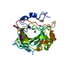
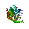
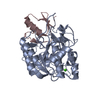
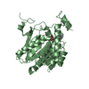
 Homo sapiens (human)
 X-RAY DIFFRACTION /
  Authors
 Citation
 Structure visualization
 Downloads & links
 Other downloads
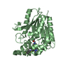


 PDBj
PDBj

 Assembly


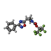
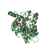
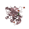
 Mass: 395.257 Da / Num. of mol.: 2 / Source method: obtained synthetically / Formula: C15H11F6N3O3 / Feature type: SUBJECT OF INVESTIGATION
 Mass: 92.094 Da / Num. of mol.: 3 / Source method: obtained synthetically / Formula: C3H8O3
 Mass: 18.015 Da / Num. of mol.: 409 / Source method: isolated from a natural source / Formula: H2O
 Sample preparation
 / Beamline: 17-ID / Wavelength: 1 Å
 Processing