 Movie
Movie Controller


+ Open data

- Basic information
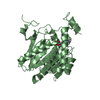
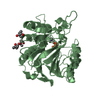
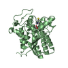
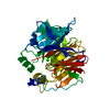
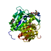
| Entry | Database: PDB / ID: 3jw8 | ||||||
|---|---|---|---|---|---|---|---|
| Title | Crystal structure of human mono-glyceride lipase | ||||||
 Components Components | MGLL protein | ||||||
 Keywords Keywords | HYDROLASE / alpha-beta hydrolase | ||||||
| Function / homology |  Function and homology information Function and homology informationArachidonate production from DAG / Acyl chain remodeling of DAG and TAG / acylglycerol catabolic process / acylglycerol lipase / monoacylglycerol catabolic process / regulation of endocannabinoid signaling pathway / monoacylglycerol lipase activity / triglyceride catabolic process / arachidonate metabolic process / regulation of sensory perception of pain ...Arachidonate production from DAG / Acyl chain remodeling of DAG and TAG / acylglycerol catabolic process / acylglycerol lipase / monoacylglycerol catabolic process / regulation of endocannabinoid signaling pathway / monoacylglycerol lipase activity / triglyceride catabolic process / arachidonate metabolic process / regulation of sensory perception of pain / phosphatidylcholine lysophospholipase A1 activity / Triglyceride catabolism / regulation of signal transduction / lipid metabolic process / fatty acid biosynthetic process / MLL4 and MLL3 complexes regulate expression of PPARG target genes in adipogenesis and hepatic steatosis / regulation of inflammatory response / inflammatory response / endoplasmic reticulum membrane / protein homodimerization activity / membrane / plasma membrane / cytosol Similarity search - Function | ||||||
| Biological species |  Homo sapiens (human) Homo sapiens (human) | ||||||
| Method |  X-RAY DIFFRACTION / SYNCHROTRON / MAD / Resolution: 2.1 Å X-RAY DIFFRACTION / SYNCHROTRON / MAD / Resolution: 2.1 Å | ||||||
 Authors Authors | Bertrand, T. / Auge, F. / Houtmann, J. / Rak, A. / Vallee, F. / Mikol, V. / Berne, P.F. / Michot, N. / Cheuret, D. / Hoornaert, C. / Mathieu, M. | ||||||
 Citation Citation | Journal: J.Mol.Biol. / Year: 2010 Title: Structural basis for human monoglyceride lipase inhibition. Authors: Bertrand, T. / Auge, F. / Houtmann, J. / Rak, A. / Vallee, F. / Mikol, V. / Berne, P.F. / Michot, N. / Cheuret, D. / Hoornaert, C. / Mathieu, M. | ||||||
| History |
|
- Structure visualization
| Structure viewer | Molecule: MolmilJmol/JSmol |
|---|
- Downloads & links
-Download
| PDBx/mmCIF format | 3jw8.cif.gz | 136.6 KB | Display | PDBx/mmCIF format |
|---|---|---|---|---|
| PDB format | pdb3jw8.ent.gz | 106.2 KB | Display | PDB format |
| PDBx/mmJSON format | 3jw8.json.gz | Tree view | PDBx/mmJSON format | |
| Others |  Other downloads Other downloads |
-Validation report
| Arichive directory | https://data.pdbj.org/pub/pdb/validation_reports/jw/3jw8ftp://data.pdbj.org/pub/pdb/validation_reports/jw/3jw8 | HTTPS FTP |
|---|
-Related structure data


-Links
 PDBj
PDBj

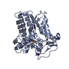
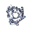
- Assembly
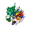
| Deposited unit | 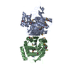
| ||||||||
|---|---|---|---|---|---|---|---|---|---|
| 1 | 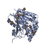
| ||||||||
| 2 | 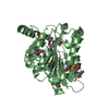
| ||||||||
| 3 | 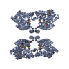
| ||||||||
| 4 | 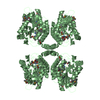
| ||||||||
| Unit cell |
|
-Components
| #1: Protein | Mass: 35808.355 Da / Num. of mol.: 2 Source method: isolated from a genetically manipulated source Source: (gene. exp.) Homo sapiens (human) / Gene: MGLL, hCG_40840 / Plasmid: pET28 / Production host:  References: UniProt: Q6IBG9, UniProt: Q99685*PLUS, acylglycerol lipase #2: Chemical |   Mass: 118.174 Da / Num. of mol.: 2 / Source method: obtained synthetically / Formula: C6H14O2 / Comment: precipitant*YM Mass: 118.174 Da / Num. of mol.: 2 / Source method: obtained synthetically / Formula: C6H14O2 / Comment: precipitant*YM#3: Chemical | ChemComp-MRD / (   Mass: 118.174 Da / Num. of mol.: 5 / Source method: obtained synthetically / Formula: C6H14O2 / Comment: precipitant*YM Mass: 118.174 Da / Num. of mol.: 5 / Source method: obtained synthetically / Formula: C6H14O2 / Comment: precipitant*YM#4: Water | ChemComp-HOH / |  Mass: 18.015 Da / Num. of mol.: 532 / Source method: isolated from a natural source / Formula: H2O Mass: 18.015 Da / Num. of mol.: 532 / Source method: isolated from a natural source / Formula: H2OHas protein modification | Y | |
|---|
-Experimental details
-Experiment
| Experiment | Method: X-RAY DIFFRACTION / Number of used crystals: 1 |
|---|
- Sample preparation
| Crystal | Density Matthews: 2.72 Å3/Da / Density % sol: 54.82 % |
|---|---|
| Crystal grow | Temperature: 277 K / Method: vapor diffusion, hanging drop / pH: 6 Details: 50mM MES, 40% MPD, pH 6.0, VAPOR DIFFUSION, HANGING DROP, temperature 277K |
-Data collection
| Diffraction | Mean temperature: 100 K | ||||||||||||
|---|---|---|---|---|---|---|---|---|---|---|---|---|---|
| Diffraction source | Source: SYNCHROTRON / Site: ESRF  / Beamline: ID23-1 / Wavelength: 0.9797, 0.9799, 1.000 / Beamline: ID23-1 / Wavelength: 0.9797, 0.9799, 1.000 | ||||||||||||
| Detector | Type: ADSC QUANTUM 315r / Detector: CCD / Date: Jan 28, 2008 | ||||||||||||
| Radiation | Monochromator: Si 111 channel / Protocol: MAD / Monochromatic (M) / Laue (L): M / Scattering type: x-ray | ||||||||||||
| Radiation wavelength |
| ||||||||||||
| Reflection | Resolution: 2.1→73.9 Å / Num. all: 44315 / Num. obs: 44315 / % possible obs: 97.8 % / Observed criterion σ(F): 0 / Observed criterion σ(I): 0 / Redundancy: 3.6 % / Biso Wilson estimate: 27.56 Å2 / Rsym value: 0.056 / Net I/σ(I): 18.4 | ||||||||||||
| Reflection shell | Resolution: 2.1→2.21 Å / Redundancy: 3.7 % / Mean I/σ(I) obs: 6.5 / Rsym value: 0.149 / % possible all: 97.4 |
- Processing
| Software |
| ||||||||||||||||||||||||||||||||||||||||||||||||||||||||||||
|---|---|---|---|---|---|---|---|---|---|---|---|---|---|---|---|---|---|---|---|---|---|---|---|---|---|---|---|---|---|---|---|---|---|---|---|---|---|---|---|---|---|---|---|---|---|---|---|---|---|---|---|---|---|---|---|---|---|---|---|---|---|
| Refinement | Method to determine structure: MAD / Resolution: 2.1→17.21 Å / Cross valid method: THROUGHOUT / σ(F): 0 / Stereochemistry target values: Engh & Huber
| ||||||||||||||||||||||||||||||||||||||||||||||||||||||||||||
| Displacement parameters | Biso mean: 27.77 Å2
| ||||||||||||||||||||||||||||||||||||||||||||||||||||||||||||
| Refine analyze | Luzzati coordinate error obs: 0.175 Å | ||||||||||||||||||||||||||||||||||||||||||||||||||||||||||||
| Refinement step | Cycle: LAST / Resolution: 2.1→17.21 Å
| ||||||||||||||||||||||||||||||||||||||||||||||||||||||||||||
| Refine LS restraints |
| ||||||||||||||||||||||||||||||||||||||||||||||||||||||||||||
| LS refinement shell | Resolution: 2.1→2.15 Å / Total num. of bins used: 20
|
