 Movie
Movie Controller


+ Open data

- Basic information
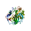
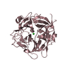
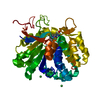
| Entry | Database: PDB / ID: 2vef | ||||||
|---|---|---|---|---|---|---|---|
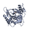
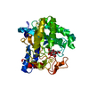
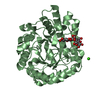
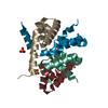
| Title | Dihydropteroate synthase from Streptococcus pneumoniae | ||||||
 Components Components | DIHYDROPTEROATE SYNTHASE | ||||||
 Keywords Keywords | TRANSFERASE / ANTIBIOTIC RESISTANCE / FOLATE BIOSYNTHESIS | ||||||
| Function / homology |  Function and homology information Function and homology informationdihydropteroate synthase / dihydropteroate synthase activity / folic acid biosynthetic process / tetrahydrofolate biosynthetic process / response to antibiotic / metal ion binding / cytosol Similarity search - Function | ||||||
| Biological species |   STREPTOCOCCUS PNEUMONIAE (bacteria) STREPTOCOCCUS PNEUMONIAE (bacteria) | ||||||
| Method |  X-RAY DIFFRACTION / SYNCHROTRON / SAD / Resolution: 1.8 Å X-RAY DIFFRACTION / SYNCHROTRON / SAD / Resolution: 1.8 Å | ||||||
 Authors Authors | Derrick, J.P. / Levy, C. | ||||||
 Citation Citation | Journal: Biochem.J. / Year: 2008 Title: Dihydropteroate Synthase from Streptococcus Pneumoniae: Structure, Ligand Recognition and Mechanism of Sulfonamide Resistance. Authors: Levy, C. / Minnis, D. / Derrick, J.P. | ||||||
| History |
|
- Structure visualization
| Structure viewer | Molecule: MolmilJmol/JSmol |
|---|
- Downloads & links
-Download
| PDBx/mmCIF format | 2vef.cif.gz | 126.8 KB | Display | PDBx/mmCIF format |
|---|---|---|---|---|
| PDB format | pdb2vef.ent.gz | 100.5 KB | Display | PDB format |
| PDBx/mmJSON format | 2vef.json.gz | Tree view | PDBx/mmJSON format | |
| Others |  Other downloads Other downloads |
-Validation report
| Arichive directory | https://data.pdbj.org/pub/pdb/validation_reports/ve/2vefftp://data.pdbj.org/pub/pdb/validation_reports/ve/2vef | HTTPS FTP |
|---|
-Related structure data


-Links
 PDBj
PDBj- Assembly
| Deposited unit | 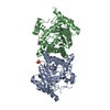
| ||||||||
|---|---|---|---|---|---|---|---|---|---|
| 1 | 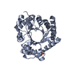
| ||||||||
| 2 | 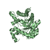
| ||||||||
| Unit cell |
| ||||||||
| Noncrystallographic symmetry (NCS) | NCS oper: (Code: given Matrix: (-0.97, -0.194, 0.148), Vector: |
-Components
| #1: Protein | Mass: 34457.195 Da / Num. of mol.: 2 Source method: isolated from a genetically manipulated source Source: (gene. exp.) STREPTOCOCCUS PNEUMONIAE (bacteria) / Production host: #2: Chemical | ChemComp-PO4 / |   Mass: 94.971 Da / Num. of mol.: 1 / Source method: obtained synthetically / Formula: PO4 Mass: 94.971 Da / Num. of mol.: 1 / Source method: obtained synthetically / Formula: PO4#3: Water | ChemComp-HOH / |  Mass: 18.015 Da / Num. of mol.: 518 / Source method: isolated from a natural source / Formula: H2O Mass: 18.015 Da / Num. of mol.: 518 / Source method: isolated from a natural source / Formula: H2OSequence details | DIPEPTIDE 'IE' REMOVED FROM THE SEQUENCE AT POSITION 65, AS THIS CORRESPONDS TO THE SULPHONAMIDE ...DIPEPTIDE 'IE' REMOVED FROM THE SEQUENCE AT POSITION 65, AS THIS CORRESPOND | |
|---|
-Experimental details
-Experiment
| Experiment | Method: X-RAY DIFFRACTION / Number of used crystals: 1 |
|---|
- Sample preparation
| Crystal | Density Matthews: 2.2 Å3/Da / Density % sol: 44.1 % / Description: NONE |
|---|---|
| Crystal grow | Details: 0.2 M AMMONIUM IODIDE, 20% (W/V) POLYETHYLENE GLYCOL 3,350 |
-Data collection
| Diffraction | Mean temperature: 100 K |
|---|---|
| Diffraction source | Source: SYNCHROTRON / Site: SRS  / Beamline: PX14.1 / Wavelength: 1.488 / Beamline: PX14.1 / Wavelength: 1.488 |
| Detector | Type: ADSC CCD / Detector: CCD |
| Radiation | Protocol: SINGLE WAVELENGTH / Monochromatic (M) / Laue (L): M / Scattering type: x-ray |
| Radiation wavelength | Wavelength: 1.488 Å / Relative weight: 1 |
| Reflection | Resolution: 1.8→55 Å / Num. obs: 65291 / % possible obs: 99 % / Observed criterion σ(I): 4 / Redundancy: 3.43 % / Rmerge(I) obs: 0.05 / Net I/σ(I): 12.7 |
| Reflection shell | Resolution: 1.8→1.9 Å / Redundancy: 2.89 % / Rmerge(I) obs: 0.21 / Mean I/σ(I) obs: 4.5 / % possible all: 93.9 |
- Processing
| Software |
| ||||||||||||||||||||||||||||||||||||||||||||||||||||||||||||||||||||||||||||||||||||||||||||||||||||||||||||||||||||||||||||||||||||||||||||||||||||||||||||||||||||||||||||||||||||||
|---|---|---|---|---|---|---|---|---|---|---|---|---|---|---|---|---|---|---|---|---|---|---|---|---|---|---|---|---|---|---|---|---|---|---|---|---|---|---|---|---|---|---|---|---|---|---|---|---|---|---|---|---|---|---|---|---|---|---|---|---|---|---|---|---|---|---|---|---|---|---|---|---|---|---|---|---|---|---|---|---|---|---|---|---|---|---|---|---|---|---|---|---|---|---|---|---|---|---|---|---|---|---|---|---|---|---|---|---|---|---|---|---|---|---|---|---|---|---|---|---|---|---|---|---|---|---|---|---|---|---|---|---|---|---|---|---|---|---|---|---|---|---|---|---|---|---|---|---|---|---|---|---|---|---|---|---|---|---|---|---|---|---|---|---|---|---|---|---|---|---|---|---|---|---|---|---|---|---|---|---|---|---|---|
| Refinement | Method to determine structure: SAD Starting model: NONE Resolution: 1.8→45.18 Å / Cor.coef. Fo:Fc: 0.951 / Cor.coef. Fo:Fc free: 0.928 / SU B: 5.816 / SU ML: 0.098 / TLS residual ADP flag: UNVERIFIED / Cross valid method: THROUGHOUT / ESU R: 0.139 / ESU R Free: 0.136 / Stereochemistry target values: MAXIMUM LIKELIHOOD / Details: HYDROGENS HAVE BEEN ADDED IN THE RIDING POSITIONS.
| ||||||||||||||||||||||||||||||||||||||||||||||||||||||||||||||||||||||||||||||||||||||||||||||||||||||||||||||||||||||||||||||||||||||||||||||||||||||||||||||||||||||||||||||||||||||
| Solvent computation | Ion probe radii: 0.8 Å / Shrinkage radii: 0.8 Å / VDW probe radii: 1.4 Å / Solvent model: MASK | ||||||||||||||||||||||||||||||||||||||||||||||||||||||||||||||||||||||||||||||||||||||||||||||||||||||||||||||||||||||||||||||||||||||||||||||||||||||||||||||||||||||||||||||||||||||
| Displacement parameters | Biso mean: 14 Å2
| ||||||||||||||||||||||||||||||||||||||||||||||||||||||||||||||||||||||||||||||||||||||||||||||||||||||||||||||||||||||||||||||||||||||||||||||||||||||||||||||||||||||||||||||||||||||
| Refinement step | Cycle: LAST / Resolution: 1.8→45.18 Å
| ||||||||||||||||||||||||||||||||||||||||||||||||||||||||||||||||||||||||||||||||||||||||||||||||||||||||||||||||||||||||||||||||||||||||||||||||||||||||||||||||||||||||||||||||||||||
| Refine LS restraints |
|