- PDB-6ekm: Crystal structure of mammalian Rev7 in complex with human Rev3 se... -
+
Open data
ID or keywords:
Loading...
-
Basic information
Entry
Database: PDB / ID: 6ekm
Title
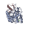
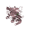
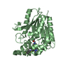
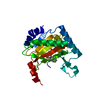
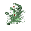
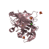
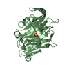
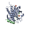
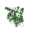
Crystal structure of mammalian Rev7 in complex with human Rev3 second binding site
Components
DNA polymerase zeta catalytic subunit
Mitotic spindle assembly checkpoint protein MAD2B
Keywords
REPLICATION / Rev7 / Mad2L2 / DNA replication / Rev3 / DNA polymerase zeta
Function / homology
Function and homology information
somatic diversification of immunoglobulins involved in immune response / DNA damage response, signal transduction resulting in transcription / Translesion synthesis by REV1 / Translesion synthesis by POLK / Translesion synthesis by POLI / negative regulation of ubiquitin protein ligase activity / zeta DNA polymerase complex / anaphase-promoting complex / positive regulation of extracellular matrix assembly / positive regulation of isotype switching ...somatic diversification of immunoglobulins involved in immune response / DNA damage response, signal transduction resulting in transcription / Translesion synthesis by REV1 / Translesion synthesis by POLK / Translesion synthesis by POLI / negative regulation of ubiquitin protein ligase activity / zeta DNA polymerase complex / anaphase-promoting complex / positive regulation of extracellular matrix assembly / positive regulation of isotype switching / : / JUN kinase binding / negative regulation of cell-cell adhesion mediated by cadherin / negative regulation of epithelial to mesenchymal transition / positive regulation of double-strand break repair via nonhomologous end joining / DNA biosynthetic process / telomere maintenance in response to DNA damage / positive regulation of peptidyl-serine phosphorylation / error-prone translesion synthesis / positive regulation of epithelial to mesenchymal transition / negative regulation of double-strand break repair via homologous recombination / site of DNA damage / translesion synthesis / actin filament organization / Translesion synthesis by REV1 / Translesion synthesis by POLK / Translesion synthesis by POLI / negative regulation of canonical Wnt signaling pathway / regulation of cell growth / negative regulation of protein catabolic process / double-strand break repair via homologous recombination / DNA-templated DNA replication / spindle / transcription corepressor activity / double-strand break repair / site of double-strand break / 4 iron, 4 sulfur cluster binding / DNA-directed DNA polymerase / RNA polymerase II-specific DNA-binding transcription factor binding / DNA-directed DNA polymerase activity / cell division / nucleotide binding / DNA repair / positive regulation of gene expression / nucleolus / positive regulation of DNA-templated transcription / chromatin / negative regulation of transcription by RNA polymerase II / DNA binding / nucleoplasm / zinc ion binding / nucleus / cytoplasm Similarity search - Function
Domain of unknown function DUF4683 / Domain of unknown function (DUF4683) / DNA polymerase zeta catalytic subunit / : / DNA polymerase zeta catalytic subunit, N-terminal / C4-type zinc-finger of DNA polymerase delta / : / C4-type zinc-finger of DNA polymerase delta / DNA polymerase delta catalytic subunit-like, N-terminal domain / Mad2-like ...Domain of unknown function DUF4683 / Domain of unknown function (DUF4683) / DNA polymerase zeta catalytic subunit / : / DNA polymerase zeta catalytic subunit, N-terminal / C4-type zinc-finger of DNA polymerase delta / : / C4-type zinc-finger of DNA polymerase delta / DNA polymerase delta catalytic subunit-like, N-terminal domain / Mad2-like / HORMA domain / HORMA domain / HORMA domain profile. / HORMA domain superfamily / DNA polymerase family B, thumb domain / DNA-directed DNA polymerase, family B, multifunctional domain / DNA-directed DNA polymerase, family B, conserved site / DNA polymerase family B signature. / DNA polymerase family B / DNA polymerase family B, exonuclease domain / DNA-directed DNA polymerase, family B, exonuclease domain / DNA polymerase, palm domain superfamily / DNA polymerase type-B family / DNA-directed DNA polymerase, family B / Ribonuclease H superfamily / Ribonuclease H-like superfamily / DNA/RNA polymerase superfamily Similarity search - Domain/homology
DNA polymerase zeta catalytic subunit / Mitotic spindle assembly checkpoint protein MAD2B Similarity search - Component
Resolution: 2.76→58.4 Å / Cor.coef. Fo:Fc: 0.923 / Cor.coef. Fo:Fc free: 0.89 / Cross valid method: THROUGHOUT / ESU R Free: 0.408 / Details: HYDROGENS HAVE BEEN ADDED IN THE RIDING POSITIONS
Rfactor
Num. reflection
% reflection
Selection details
Rfree
0.25843
298
4.9 %
RANDOM
Rwork
0.23009
-
-
-
obs
0.23152
5820
99.5 %
-
Solvent computation
Ion probe radii: 0.8 Å / Shrinkage radii: 0.8 Å / VDW probe radii: 1.2 Å
 Movie
Movie Controller

 Yorodumi
 Open data
 Basic information
 Components
 Keywords
 Function and homology information
 Homo sapiens (human)
 X-RAY DIFFRACTION /
  Authors
 Switzerland, 1items
  Citation
 Structure visualization
 Downloads & links
 Other downloads
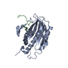

 PDBj
PDBj


 Assembly

 Mass: 18.015 Da / Num. of mol.: 21 / Source method: isolated from a natural source / Formula: H2O
 Sample preparation
 Processing