Arachidonate production from DAG / Acyl chain remodeling of DAG and TAG / acylglycerol catabolic process / acylglycerol lipase / monoacylglycerol catabolic process / regulation of endocannabinoid signaling pathway / monoacylglycerol lipase activity / triglyceride catabolic process / arachidonate metabolic process / regulation of sensory perception of pain ...Arachidonate production from DAG / Acyl chain remodeling of DAG and TAG / acylglycerol catabolic process / acylglycerol lipase / monoacylglycerol catabolic process / regulation of endocannabinoid signaling pathway / monoacylglycerol lipase activity / triglyceride catabolic process / arachidonate metabolic process / regulation of sensory perception of pain / phosphatidylcholine lysophospholipase A1 activity / Triglyceride catabolism / regulation of signal transduction / lipid metabolic process / fatty acid biosynthetic process / MLL4 and MLL3 complexes regulate expression of PPARG target genes in adipogenesis and hepatic steatosis / regulation of inflammatory response / inflammatory response / endoplasmic reticulum membrane / protein homodimerization activity / membrane / plasma membrane / cytosol Similarity search - Function
Resolution: 1.35→40 Å / Cor.coef. Fo:Fc: 0.976 / Cor.coef. Fo:Fc free: 0.971 / SU B: 1.479 / SU ML: 0.029 / Cross valid method: THROUGHOUT / ESU R: 0.042 / ESU R Free: 0.042 / Details: HYDROGENS HAVE BEEN ADDED IN THE RIDING POSITIONS
Rfactor
Num. reflection
% reflection
Selection details
Rfree
0.16008
3916
5 %
RANDOM
Rwork
0.14693
-
-
-
obs
0.14758
74657
97.83 %
-
Solvent computation
Ion probe radii: 0.8 Å / Shrinkage radii: 0.8 Å / VDW probe radii: 1.2 Å
 Movie
Movie Controller

 Yorodumi
 Open data
 Basic information
 Components
 Keywords
 Function and homology information
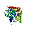
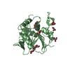
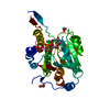
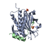
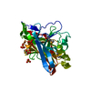
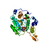
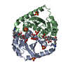
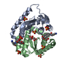
 Homo sapiens (human)
 X-RAY DIFFRACTION /
  Authors
 Citation
 Structure visualization
 Downloads & links
 Other downloads


 PDBj
PDBj

 Assembly
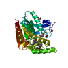

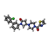


 Mass: 466.983 Da / Num. of mol.: 1 / Source method: obtained synthetically / Formula: C24H23ClN4O2S
 Mass: 194.226 Da / Num. of mol.: 1 / Source method: obtained synthetically / Formula: C8H18O5 / Comment: precipitant*YM
 Mass: 62.068 Da / Num. of mol.: 8 / Source method: obtained synthetically / Formula: C2H6O2
 Mass: 35.453 Da / Num. of mol.: 1 / Source method: obtained synthetically / Formula: Cl
 Sample preparation
 / Beamline: 5.0.2 / Wavelength: 1 Å
 Processing