 Movie
Movie Controller

[English] 日本語
 Yorodumi
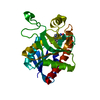
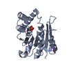
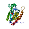
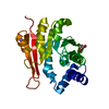
- PDB-6aw4: 1.50A resolution structure of catechol O-methyltransferase (COMT)... -
+ Open data

- Basic information
| Entry | Database: PDB / ID: 6aw4 | ||||||
|---|---|---|---|---|---|---|---|
| Title | 1.50A resolution structure of catechol O-methyltransferase (COMT) from Nannospalax galili | ||||||
 Components Components | Catechol O-methyltransferase | ||||||
 Keywords Keywords | TRANSFERASE / Spalax / COMT / S-adenosylmethionine binding | ||||||
| Function / homology |  Function and homology information Function and homology informationcatecholamine catabolic process / catechol O-methyltransferase activity / catechol O-methyltransferase / developmental process / dopamine metabolic process / methylation / axon / dendrite / magnesium ion binding / plasma membrane Similarity search - Function | ||||||
| Biological species |  Nannospalax galili (Upper Galilee mountains blind mole rat) Nannospalax galili (Upper Galilee mountains blind mole rat) | ||||||
| Method |  X-RAY DIFFRACTION / SYNCHROTRON / MOLECULAR REPLACEMENT / molecular replacement / Resolution: 1.5 Å X-RAY DIFFRACTION / SYNCHROTRON / MOLECULAR REPLACEMENT / molecular replacement / Resolution: 1.5 Å | ||||||
 Authors Authors | Lovell, S. / Mehzabeen, N. / Battaile, K.P. / Deng, Y. / Hanzlik, R.P. / Shams, I. / Moskovitz, J. | ||||||
| Funding support |  United States, 1items United States, 1items
| ||||||
 Citation Citation | Journal: To be published Title: Crystal structure of the catechol-o-methyl transferase (COMT) enzyme of the subterranean mole rat (Spalax) and the effect of L136M substitution Authors: Deng, Y. / Lovell, S. / Mehzabeen, N. / Battaile, K.P. / Hanzlik, R.P. / Shams, I. / Moskovitz, J. | ||||||
| History |
|
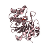
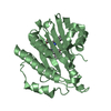
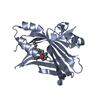
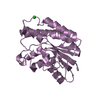
- Structure visualization
| Structure viewer | Molecule: MolmilJmol/JSmol |
|---|
- Downloads & links
-Download
| PDBx/mmCIF format | 6aw4.cif.gz | 65 KB | Display | PDBx/mmCIF format |
|---|---|---|---|---|
| PDB format | pdb6aw4.ent.gz | 44.7 KB | Display | PDB format |
| PDBx/mmJSON format | 6aw4.json.gz | Tree view | PDBx/mmJSON format | |
| Others |  Other downloads Other downloads |
-Validation report
| Arichive directory | https://data.pdbj.org/pub/pdb/validation_reports/aw/6aw4ftp://data.pdbj.org/pub/pdb/validation_reports/aw/6aw4 | HTTPS FTP |
|---|
-Related structure data
| Related structure data |  6aw5C  6aw6C  6aw7C  6aw8C  6aw9C  2zlbS S: Starting model for refinement C: citing same article ( |
|---|---|
| Similar structure data |


-Links
 PDBj
PDBj- Assembly
| Deposited unit | 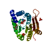
| ||||||||
|---|---|---|---|---|---|---|---|---|---|
| 1 |
| ||||||||
| Unit cell |
| ||||||||
| Components on special symmetry positions |
|
-Components
-Protein , 1 types, 1 molecules A
| #1: Protein | Mass: 25717.373 Da / Num. of mol.: 1 / Fragment: M43-P262 Source method: isolated from a genetically manipulated source Source: (gene. exp.) Nannospalax galili (Upper Galilee mountains blind mole rat)Plasmid: pET28a / Production host:  |
|---|
-Non-polymers , 5 types, 215 molecules 


| #2: Chemical |  Mass: 92.094 Da / Num. of mol.: 2 / Source method: obtained synthetically / Formula: C3H8O3 Mass: 92.094 Da / Num. of mol.: 2 / Source method: obtained synthetically / Formula: C3H8O3#3: Chemical | ChemComp-NA / |  Mass: 22.990 Da / Num. of mol.: 1 / Source method: obtained synthetically / Formula: Na Mass: 22.990 Da / Num. of mol.: 1 / Source method: obtained synthetically / Formula: Na#4: Chemical | ChemComp-TRS / |  Mass: 122.143 Da / Num. of mol.: 1 / Source method: obtained synthetically / Formula: C4H12NO3 / Comment: pH buffer*YM Mass: 122.143 Da / Num. of mol.: 1 / Source method: obtained synthetically / Formula: C4H12NO3 / Comment: pH buffer*YM#5: Chemical | ChemComp-PO4 /  Mass: 94.971 Da / Num. of mol.: 4 / Source method: obtained synthetically / Formula: PO4 Mass: 94.971 Da / Num. of mol.: 4 / Source method: obtained synthetically / Formula: PO4#6: Water | ChemComp-HOH / | Mass: 18.015 Da / Num. of mol.: 207 / Source method: isolated from a natural source / Formula: H2O |
|---|
-Experimental details
-Experiment
| Experiment | Method: X-RAY DIFFRACTION / Number of used crystals: 1 |
|---|
- Sample preparation
| Crystal | Density Matthews: 3.83 Å3/Da / Density % sol: 67.86 % / Mosaicity: 0.09 ° |
|---|---|
| Crystal grow | Temperature: 293 K / Method: vapor diffusion, sitting drop / pH: 5 Details: 1.8 M Sodium phosphate monobasic monohydrate, Potassium phosphate dibasic |
-Data collection
| Diffraction | Mean temperature: 100 K |
|---|---|
| Diffraction source | Source: SYNCHROTRON / Site: APS / Beamline: 17-ID / Wavelength: 1 Å |
| Detector | Type: DECTRIS PILATUS 6M / Detector: PIXEL / Date: Oct 28, 2015 |
| Radiation | Protocol: SINGLE WAVELENGTH / Monochromatic (M) / Laue (L): M / Scattering type: x-ray |
| Radiation wavelength | Wavelength: 1 Å / Relative weight: 1 |
| Reflection | Resolution: 1.5→47.38 Å / Num. obs: 62014 / % possible obs: 100 % / Redundancy: 10.2 % / Biso Wilson estimate: 21.32 Å2 / CC1/2: 0.999 / Rmerge(I) obs: 0.053 / Net I/σ(I): 20.9 |
| Reflection shell | Resolution: 1.5→1.53 Å / Redundancy: 10.1 % / Rmerge(I) obs: 1.431 / Num. measured all: 30834 / Num. unique all: 3066 / CC1/2: 0.663 / Net I/σ(I) obs: 1.9 / % possible all: 100 |
-Phasing
| Phasing | Method: molecular replacement | |||||||||
|---|---|---|---|---|---|---|---|---|---|---|
| Phasing MR | Model details: Phaser MODE: MR_AUTO
|
- Processing
| Software |
| ||||||||||||||||||||||||||||||||||||||||||||||||||||||||||||||||||||||||||||||||||||||||||||||||||||||||||||||||||||||||||||||||||||||||||
|---|---|---|---|---|---|---|---|---|---|---|---|---|---|---|---|---|---|---|---|---|---|---|---|---|---|---|---|---|---|---|---|---|---|---|---|---|---|---|---|---|---|---|---|---|---|---|---|---|---|---|---|---|---|---|---|---|---|---|---|---|---|---|---|---|---|---|---|---|---|---|---|---|---|---|---|---|---|---|---|---|---|---|---|---|---|---|---|---|---|---|---|---|---|---|---|---|---|---|---|---|---|---|---|---|---|---|---|---|---|---|---|---|---|---|---|---|---|---|---|---|---|---|---|---|---|---|---|---|---|---|---|---|---|---|---|---|---|---|---|
| Refinement | Method to determine structure: MOLECULAR REPLACEMENT Starting model: 2ZLB Resolution: 1.5→34.458 Å / SU ML: 0.12 / Cross valid method: FREE R-VALUE / σ(F): 1.01 / Phase error: 15.88
| ||||||||||||||||||||||||||||||||||||||||||||||||||||||||||||||||||||||||||||||||||||||||||||||||||||||||||||||||||||||||||||||||||||||||||
| Solvent computation | Shrinkage radii: 0.9 Å / VDW probe radii: 1.11 Å | ||||||||||||||||||||||||||||||||||||||||||||||||||||||||||||||||||||||||||||||||||||||||||||||||||||||||||||||||||||||||||||||||||||||||||
| Displacement parameters | Biso max: 69.57 Å2 / Biso mean: 26.9413 Å2 / Biso min: 13.73 Å2 | ||||||||||||||||||||||||||||||||||||||||||||||||||||||||||||||||||||||||||||||||||||||||||||||||||||||||||||||||||||||||||||||||||||||||||
| Refinement step | Cycle: final / Resolution: 1.5→34.458 Å
| ||||||||||||||||||||||||||||||||||||||||||||||||||||||||||||||||||||||||||||||||||||||||||||||||||||||||||||||||||||||||||||||||||||||||||
| Refine LS restraints |
| ||||||||||||||||||||||||||||||||||||||||||||||||||||||||||||||||||||||||||||||||||||||||||||||||||||||||||||||||||||||||||||||||||||||||||
| LS refinement shell | Refine-ID: X-RAY DIFFRACTION / Rfactor Rfree error: 0 / Total num. of bins used: 22 / % reflection obs: 100 %
|
