 Movie
Movie Controller

[English] 日本語
 Yorodumi
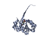
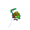
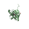
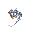
- PDB-6aw9: 2.55A resolution structure of SAH bound catechol O-methyltransfer... -
+ Open data

- Basic information
| Entry | Database: PDB / ID: 6aw9 | ||||||
|---|---|---|---|---|---|---|---|
| Title | 2.55A resolution structure of SAH bound catechol O-methyltransferase (COMT) L136M from Nannospalax galili | ||||||
 Components Components | Catechol O-methyltransferase | ||||||
 Keywords Keywords | TRANSFERASE / Spalax / COMT / S-adenosylmethionine binding | ||||||
| Function / homology |  Function and homology information Function and homology informationcatecholamine catabolic process / catechol O-methyltransferase activity / catechol O-methyltransferase / developmental process / dopamine metabolic process / methylation / axon / dendrite / magnesium ion binding / plasma membrane Similarity search - Function | ||||||
| Biological species |  Nannospalax galili (Upper Galilee mountains blind mole rat) Nannospalax galili (Upper Galilee mountains blind mole rat) | ||||||
| Method |  X-RAY DIFFRACTION / SYNCHROTRON / MOLECULAR REPLACEMENT / molecular replacement / Resolution: 2.55 Å X-RAY DIFFRACTION / SYNCHROTRON / MOLECULAR REPLACEMENT / molecular replacement / Resolution: 2.55 Å | ||||||
 Authors Authors | Lovell, S. / Mehzabeen, N. / Battaile, K.P. / Deng, Y. / Hanzlik, R.P. / Shams, I. / Moskovitz, J. | ||||||
| Funding support |  United States, 1items United States, 1items
| ||||||
 Citation Citation | Journal: To be published Title: Crystal structure of the catechol-o-methyl transferase (COMT) enzyme of the subterranean mole rat (Spalax) and the effect of L136M substitution Authors: Deng, Y. / Lovell, S. / Mehzabeen, N. / Battaile, K.P. / Hanzlik, R.P. / Shams, I. / Moskovitz, J. | ||||||
| History |
|
- Structure visualization
| Structure viewer | Molecule: MolmilJmol/JSmol |
|---|
- Downloads & links
-Download
| PDBx/mmCIF format | 6aw9.cif.gz | 139.3 KB | Display | PDBx/mmCIF format |
|---|---|---|---|---|
| PDB format | pdb6aw9.ent.gz | 106.3 KB | Display | PDB format |
| PDBx/mmJSON format | 6aw9.json.gz | Tree view | PDBx/mmJSON format | |
| Others |  Other downloads Other downloads |
-Validation report
| Arichive directory | https://data.pdbj.org/pub/pdb/validation_reports/aw/6aw9ftp://data.pdbj.org/pub/pdb/validation_reports/aw/6aw9 | HTTPS FTP |
|---|
-Related structure data
| Related structure data |  6aw4C  6aw5C  6aw6C  6aw7C  6aw8C  2zlbS S: Starting model for refinement C: citing same article ( |
|---|---|
| Similar structure data |


-Links
 PDBj
PDBj- Assembly
| Deposited unit | 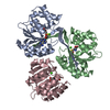
| ||||||||
|---|---|---|---|---|---|---|---|---|---|
| 1 | 
| ||||||||
| 2 | 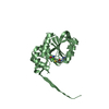
| ||||||||
| 3 | 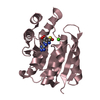
| ||||||||
| Unit cell |
|
-Components
| #1: Protein | Mass: 25735.410 Da / Num. of mol.: 3 / Fragment: M43-P262 / Mutation: L136M Source method: isolated from a genetically manipulated source Source: (gene. exp.) Nannospalax galili (Upper Galilee mountains blind mole rat)Plasmid: pET28a / Production host:  #2: Chemical |   Mass: 40.078 Da / Num. of mol.: 3 / Source method: obtained synthetically / Formula: Ca Mass: 40.078 Da / Num. of mol.: 3 / Source method: obtained synthetically / Formula: Ca#3: Chemical |   Type: L-peptide linking / Mass: 384.411 Da / Num. of mol.: 3 / Source method: obtained synthetically / Formula: C14H20N6O5S Type: L-peptide linking / Mass: 384.411 Da / Num. of mol.: 3 / Source method: obtained synthetically / Formula: C14H20N6O5S#4: Water | ChemComp-HOH / |  Mass: 18.015 Da / Num. of mol.: 34 / Source method: isolated from a natural source / Formula: H2O Mass: 18.015 Da / Num. of mol.: 34 / Source method: isolated from a natural source / Formula: H2O |
|---|
-Experimental details
-Experiment
| Experiment | Method: X-RAY DIFFRACTION / Number of used crystals: 1 |
|---|
- Sample preparation
| Crystal | Density Matthews: 2.64 Å3/Da / Density % sol: 53.34 % / Mosaicity: 0.14 ° |
|---|---|
| Crystal grow | Temperature: 293 K / Method: vapor diffusion, sitting drop / pH: 6.5 Details: 14.4 % (w/v) PEG 8000, 80 mM sodium cacodylate, 160 mM calcium chloride, 20% glycerol |
-Data collection
| Diffraction | Mean temperature: 100 K |
|---|---|
| Diffraction source | Source: SYNCHROTRON / Site: APS / Beamline: 17-ID / Wavelength: 1 Å |
| Detector | Type: DECTRIS PILATUS 6M / Detector: PIXEL / Date: Apr 10, 2016 |
| Radiation | Protocol: SINGLE WAVELENGTH / Monochromatic (M) / Laue (L): M / Scattering type: x-ray |
| Radiation wavelength | Wavelength: 1 Å / Relative weight: 1 |
| Reflection | Resolution: 2.55→49.49 Å / Num. obs: 27248 / % possible obs: 99.7 % / Redundancy: 6.6 % / Biso Wilson estimate: 52.98 Å2 / CC1/2: 0.998 / Rmerge(I) obs: 0.114 / Net I/σ(I): 11.7 / Num. measured all: 179065 / Scaling rejects: 0 |
| Reflection shell | Resolution: 2.55→2.66 Å / Redundancy: 6.5 % / Rmerge(I) obs: 1.2 / Num. measured all: 21344 / Num. unique all: 3260 / CC1/2: 0.791 / Net I/σ(I) obs: 1.5 / % possible all: 99.4 |
-Phasing
| Phasing | Method: molecular replacement | |||||||||
|---|---|---|---|---|---|---|---|---|---|---|
| Phasing MR | Model details: Phaser MODE: MR_AUTO
|
- Processing
| Software |
| |||||||||||||||||||||||||||||||||||||||||||||||||||||||||||||||||||||||||||||
|---|---|---|---|---|---|---|---|---|---|---|---|---|---|---|---|---|---|---|---|---|---|---|---|---|---|---|---|---|---|---|---|---|---|---|---|---|---|---|---|---|---|---|---|---|---|---|---|---|---|---|---|---|---|---|---|---|---|---|---|---|---|---|---|---|---|---|---|---|---|---|---|---|---|---|---|---|---|---|
| Refinement | Method to determine structure: MOLECULAR REPLACEMENT Starting model: 2ZLB Resolution: 2.55→44.579 Å / SU ML: 0.41 / Cross valid method: FREE R-VALUE / σ(F): 1.36 / Phase error: 32.72
| |||||||||||||||||||||||||||||||||||||||||||||||||||||||||||||||||||||||||||||
| Solvent computation | Shrinkage radii: 0.9 Å / VDW probe radii: 1.11 Å | |||||||||||||||||||||||||||||||||||||||||||||||||||||||||||||||||||||||||||||
| Displacement parameters | Biso max: 166.27 Å2 / Biso mean: 70.9638 Å2 / Biso min: 34.25 Å2 | |||||||||||||||||||||||||||||||||||||||||||||||||||||||||||||||||||||||||||||
| Refinement step | Cycle: final / Resolution: 2.55→44.579 Å
| |||||||||||||||||||||||||||||||||||||||||||||||||||||||||||||||||||||||||||||
| Refine LS restraints |
| |||||||||||||||||||||||||||||||||||||||||||||||||||||||||||||||||||||||||||||
| LS refinement shell | Refine-ID: X-RAY DIFFRACTION / Rfactor Rfree error: 0 / Total num. of bins used: 10
|
