 Movie
Movie Controller


+ Open data

- Basic information
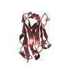
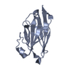
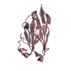
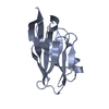
| Entry | Database: PDB / ID: 6asr | ||||||
|---|---|---|---|---|---|---|---|
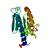
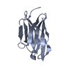
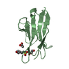
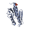
| Title | REV1 UBM2 domain complex with ubiquitin | ||||||
 Components Components |
| ||||||
 Keywords Keywords | PROTEIN BINDING / ubiquitin-binding motif / protein-protein interaction | ||||||
| Function / homology |  Function and homology information Function and homology informationdeoxycytidyl transferase activity / symbiont entry into host cell via disruption of host cell glycocalyx / error-free translesion synthesis / symbiont entry into host cell via disruption of host cell envelope / virus tail / error-prone translesion synthesis / response to UV / Translesion synthesis by REV1 / Translesion synthesis by POLK / Translesion synthesis by POLI ...deoxycytidyl transferase activity / symbiont entry into host cell via disruption of host cell glycocalyx / error-free translesion synthesis / symbiont entry into host cell via disruption of host cell envelope / virus tail / error-prone translesion synthesis / response to UV / Translesion synthesis by REV1 / Translesion synthesis by POLK / Translesion synthesis by POLI / Termination of translesion DNA synthesis / Transferases; Transferring phosphorus-containing groups; Nucleotidyltransferases / damaged DNA binding / DNA-directed DNA polymerase activity / DNA replication / nucleoplasm / metal ion binding Similarity search - Function | ||||||
| Biological species |  Homo sapiens (human) Homo sapiens (human) | ||||||
| Method |  X-RAY DIFFRACTION / SYNCHROTRON / MOLECULAR REPLACEMENT / Resolution: 2.356 Å X-RAY DIFFRACTION / SYNCHROTRON / MOLECULAR REPLACEMENT / Resolution: 2.356 Å | ||||||
 Authors Authors | Miller, D.J. | ||||||
 Citation Citation | Journal: J. Mol. Biol. / Year: 2018 Title: Structures of REV1 UBM2 Domain Complex with Ubiquitin and with a Small-Molecule that Inhibits the REV1 UBM2-Ubiquitin Interaction. Authors: Vanarotti, M. / Grace, C.R. / Miller, D.J. / Actis, M.L. / Inoue, A. / Evison, B.J. / Vaithiyalingam, S. / Singh, A.P. / McDonald, E.T. / Fujii, N. | ||||||
| History |
|
- Structure visualization
| Structure viewer | Molecule: MolmilJmol/JSmol |
|---|
- Downloads & links
-Download
| PDBx/mmCIF format | 6asr.cif.gz | 53.9 KB | Display | PDBx/mmCIF format |
|---|---|---|---|---|
| PDB format | pdb6asr.ent.gz | 38.2 KB | Display | PDB format |
| PDBx/mmJSON format | 6asr.json.gz | Tree view | PDBx/mmJSON format | |
| Others |  Other downloads Other downloads |
-Validation report
| Summary document | 6asr_validation.pdf.gz | 437 KB | Display | wwPDB validaton report |
|---|---|---|---|---|
| Full document | 6asr_full_validation.pdf.gz | 437.2 KB | Display | |
| Data in XML | 6asr_validation.xml.gz | 9.5 KB | Display | |
| Data in CIF | 6asr_validation.cif.gz | 12.3 KB | Display | |
| Arichive directory | https://data.pdbj.org/pub/pdb/validation_reports/as/6asrftp://data.pdbj.org/pub/pdb/validation_reports/as/6asr | HTTPS FTP |
-Related structure data
| Related structure data | 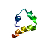 6axdC 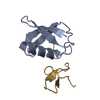 4s1zS S: Starting model for refinement C: citing same article ( |
|---|---|
| Similar structure data |


-Links
 PDBj
PDBj


- Assembly
| Deposited unit | 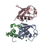
| ||||||||
|---|---|---|---|---|---|---|---|---|---|
| 1 | 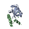
| ||||||||
| 2 | 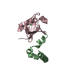
| ||||||||
| Unit cell |
| ||||||||
| Components on special symmetry positions |
|
-Components
| #1: Protein | Mass: 8576.831 Da / Num. of mol.: 2 / Fragment: residues 1-76 Source method: isolated from a genetically manipulated source Source: (gene. exp.) Homo sapiens (human) / Gene: UBB / Production host:  #2: Protein | | Mass: 5420.993 Da / Num. of mol.: 1 / Fragment: residues 998-1040 Source method: isolated from a genetically manipulated source Source: (gene. exp.) Homo sapiens (human) / Gene: REV1, REV1L / Production host: References: UniProt: Q9UBZ9, Transferases; Transferring phosphorus-containing groups; Nucleotidyltransferases #3: Chemical | ChemComp-NI /   Mass: 58.693 Da / Num. of mol.: 4 / Source method: obtained synthetically / Formula: Ni Mass: 58.693 Da / Num. of mol.: 4 / Source method: obtained synthetically / Formula: Ni#4: Water | ChemComp-HOH / |  Mass: 18.015 Da / Num. of mol.: 73 / Source method: isolated from a natural source / Formula: H2O Mass: 18.015 Da / Num. of mol.: 73 / Source method: isolated from a natural source / Formula: H2O |
|---|
-Experimental details
-Experiment
| Experiment | Method: X-RAY DIFFRACTION / Number of used crystals: 1 |
|---|
- Sample preparation
| Crystal | Density Matthews: 4.14 Å3/Da / Density % sol: 70.29 % |
|---|---|
| Crystal grow | Temperature: 291 K / Method: vapor diffusion, sitting drop / pH: 6 / Details: 0.1 M MES pH 6.0, 10 % PEG 8000 / PH range: 6 |
-Data collection
| Diffraction | Mean temperature: 100 K |
|---|---|
| Diffraction source | Source: SYNCHROTRON / Site: APS  / Beamline: 22-ID / Wavelength: 1 Å / Beamline: 22-ID / Wavelength: 1 Å |
| Detector | Type: MARMOSAIC 300 mm CCD / Detector: CCD / Date: Jul 19, 2017 |
| Radiation | Protocol: SINGLE WAVELENGTH / Monochromatic (M) / Laue (L): M / Scattering type: x-ray |
| Radiation wavelength | Wavelength: 1 Å / Relative weight: 1 |
| Reflection | Resolution: 2.35→30 Å / Num. obs: 16003 / % possible obs: 99.1 % / Observed criterion σ(I): -3 / Redundancy: 6.9 % / Rsym value: 0.106 / Net I/σ(I): 15.9 |
| Reflection shell | Resolution: 2.35→2.39 Å / Redundancy: 5 % / Num. unique obs: 781 / Rsym value: 0.532 / % possible all: 100 |
- Processing
| Software |
| |||||||||||||||||||||||||||||||||||||||||||||||||
|---|---|---|---|---|---|---|---|---|---|---|---|---|---|---|---|---|---|---|---|---|---|---|---|---|---|---|---|---|---|---|---|---|---|---|---|---|---|---|---|---|---|---|---|---|---|---|---|---|---|---|
| Refinement | Method to determine structure: MOLECULAR REPLACEMENT Starting model: 4s1z Resolution: 2.356→28.284 Å / SU ML: 0.2 / Cross valid method: THROUGHOUT / σ(F): 1.36 / Phase error: 23.05 / Stereochemistry target values: ML
| |||||||||||||||||||||||||||||||||||||||||||||||||
| Solvent computation | Shrinkage radii: 0.9 Å / VDW probe radii: 1.11 Å / Solvent model: FLAT BULK SOLVENT MODEL | |||||||||||||||||||||||||||||||||||||||||||||||||
| Refinement step | Cycle: LAST / Resolution: 2.356→28.284 Å
| |||||||||||||||||||||||||||||||||||||||||||||||||
| Refine LS restraints |
| |||||||||||||||||||||||||||||||||||||||||||||||||
| LS refinement shell |
|
