positive regulation of lyase activity / [4Fe-4S] cluster assembly / Mitochondrial iron-sulfur cluster biogenesis / Complex III assembly / iron chaperone activity / Maturation of TCA enzymes and regulation of TCA cycle / mitochondrial respiratory chain complex III assembly / Mitochondrial protein import / mitochondrial [2Fe-2S] assembly complex / response to iron ion ...positive regulation of lyase activity / [4Fe-4S] cluster assembly / Mitochondrial iron-sulfur cluster biogenesis / Complex III assembly / iron chaperone activity / Maturation of TCA enzymes and regulation of TCA cycle / mitochondrial respiratory chain complex III assembly / Mitochondrial protein import / mitochondrial [2Fe-2S] assembly complex / response to iron ion / heme biosynthetic process / [2Fe-2S] cluster assembly / iron-sulfur cluster assembly / negative regulation of release of cytochrome c from mitochondria / ferroxidase / protein autoprocessing / ferroxidase activity / ferric iron binding / protein maturation / iron ion transport / ferrous iron binding / 2 iron, 2 sulfur cluster binding / cellular response to hydrogen peroxide / enzyme activator activity / intracellular iron ion homeostasis / mitochondrial matrix / negative regulation of apoptotic process / mitochondrion / cytosol Similarity search - Function
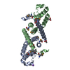
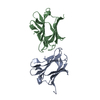
Frataxin/CyaY / Frataxin / Metal Transport, Frataxin; Chain A / Frataxin/CyaY / Frataxin conserved site / Frataxin-like domain / Frataxin family signature. / Frataxin family profile. / Frataxin-like domain / Frataxin/CyaY superfamily ...Frataxin/CyaY / Frataxin / Metal Transport, Frataxin; Chain A / Frataxin/CyaY / Frataxin conserved site / Frataxin-like domain / Frataxin family signature. / Frataxin family profile. / Frataxin-like domain / Frataxin/CyaY superfamily / 2-Layer Sandwich / Alpha Beta Similarity search - Domain/homology
In the structure databanks used in Yorodumi, some data are registered as the other names, "COVID-19 virus" and "2019-nCoV". Here are the details of the virus and the list of structure data.
Jan 31, 2019. EMDB accession codes are about to change! (news from PDBe EMDB page)
EMDB accession codes are about to change! (news from PDBe EMDB page)
The allocation of 4 digits for EMDB accession codes will soon come to an end. Whilst these codes will remain in use, new EMDB accession codes will include an additional digit and will expand incrementally as the available range of codes is exhausted. The current 4-digit format prefixed with “EMD-” (i.e. EMD-XXXX) will advance to a 5-digit format (i.e. EMD-XXXXX), and so on. It is currently estimated that the 4-digit codes will be depleted around Spring 2019, at which point the 5-digit format will come into force.
The EM Navigator/Yorodumi systems omit the EMD- prefix.
Related info.:Q: What is EMD? / ID/Accession-code notation in Yorodumi/EM Navigator
Yorodumi is a browser for structure data from EMDB, PDB, SASBDB, etc.
This page is also the successor to EM Navigator detail page, and also detail information page/front-end page for Omokage search.
The word "yorodu" (or yorozu) is an old Japanese word meaning "ten thousand". "mi" (miru) is to see.
Related info.:EMDB / PDB / SASBDB / Comparison of 3 databanks / Yorodumi Search / Aug 31, 2016. New EM Navigator & Yorodumi / Yorodumi Papers / Jmol/JSmol / Function and homology information / Changes in new EM Navigator and Yorodumi
 Movie
Movie Controller


 Open data
 Basic information
 Components
 Keywords
 Function and homology information
 Homo sapiens (human)
 X-RAY DIFFRACTION /
  Authors
 Citation
 Structure visualization
 Downloads & links
 Other downloads


 PDBj
PDBj

 Assembly
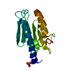


 Mass: 96.063 Da / Num. of mol.: 1 / Source method: obtained synthetically / Formula: SO4
 Mass: 18.015 Da / Num. of mol.: 102 / Source method: isolated from a natural source / Formula: H2O
 Sample preparation
 / Beamline: 23-ID-D / Wavelength: 1.03315
  Processing