National Institutes of Health/National Institute of General Medical Sciences (NIH/NIGMS)
GM81303
米国
National Institutes of Health/National Institute of General Medical Sciences (NIH/NIGMS)
GM44757
米国
National Institutes of Health/National Institute on Aging (NIH/NIA)
AG32961
米国
引用
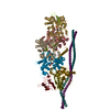
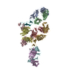
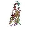
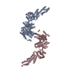
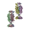
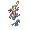
ジャーナル: Nat Commun / 年: 2017 タイトル: Structural basis for high-affinity actin binding revealed by a β-III-spectrin SCA5 missense mutation. 著者: Adam W Avery / Michael E Fealey / Fengbin Wang / Albina Orlova / Andrew R Thompson / David D Thomas / Thomas S Hays / Edward H Egelman / 要旨: Spinocerebellar ataxia type 5 (SCA5) is a neurodegenerative disease caused by mutations in the cytoskeletal protein β-III-spectrin. Previously, a SCA5 mutation resulting in a leucine-to-proline ...Spinocerebellar ataxia type 5 (SCA5) is a neurodegenerative disease caused by mutations in the cytoskeletal protein β-III-spectrin. Previously, a SCA5 mutation resulting in a leucine-to-proline substitution (L253P) in the actin-binding domain (ABD) was shown to cause a 1000-fold increase in actin-binding affinity. However, the structural basis for this increase is unknown. Here, we report a 6.9 Å cryo-EM structure of F-actin complexed with the L253P ABD. This structure, along with co-sedimentation and pulsed-EPR measurements, demonstrates that high-affinity binding caused by the CH2-localized mutation is due to opening of the two CH domains. This enables CH1 to bind actin aided by an unstructured N-terminal region that becomes α-helical upon binding. This helix is required for association with actin as truncation eliminates binding. Collectively, these results shed light on the mechanism by which β-III-spectrin, and likely similar actin-binding proteins, interact with actin, and how this mechanism can be perturbed to cause disease.
根拠: microscopy, helical filament was observed by negative staining and Cryo-EM
タイプ
名称
対称操作
数
identity operation
1_555
1
Buried area
36340 Å2
ΔGint
-117 kcal/mol
Surface area
108660 Å2
対称性
らせん対称: (回転対称性: 1 / Dyad axis: no / N subunits divisor: 1 / Num. of operations: 20 / Rise per n subunits: 27.25 Å / Rotation per n subunits: -166.87 °)
詳細
THE ASSEMBLY REPRESENTED IN THIS ENTRY HAS REGULAR HELICAL SYMMETRY WITH THE FOLLOWING PARAMETERS: ROTATION PER SUBUNIT (TWIST) = -166.87 DEGREES RISE PER SUBUNIT (HEIGHT) = 27.25 ANGSTROMS
平均露光時間: 3 sec. / 電子線照射量: 20 e/Å2 / 検出モード: INTEGRATING フィルム・検出器のモデル: FEI FALCON II (4k x 4k) 詳細: Images were stored containing seven parts, where each part represented a set of frames corresponding to a dose of ~20 electrons per Angstrom^2. The full dose image stack was used for the ...詳細: Images were stored containing seven parts, where each part represented a set of frames corresponding to a dose of ~20 electrons per Angstrom^2. The full dose image stack was used for the estimation of the CTF as well as for boxing filaments. Only the first two parts were used for the reconstruction (~5 electrons per Angstrom^2).
画像スキャン
動画フレーム数/画像: 7
-
解析
ソフトウェア
名称: PHENIX / バージョン: dev_2471: / 分類: 精密化
EMソフトウェア
ID
名称
カテゴリ
1
EMAN2
粒子像選択
2
EPU
画像取得
4
CTFFIND3
CTF補正
7
Rosetta
モデルフィッティング
9
SPIDER
初期オイラー角割当
10
SPIDER
最終オイラー角割当
12
SPIDER
3次元再構成
13
PHENIX
モデル精密化
14
Coot
モデル精密化
CTF補正
タイプ: PHASE FLIPPING AND AMPLITUDE CORRECTION
らせん対称
回転角度/サブユニット: -166.87 ° / 軸方向距離/サブユニット: 27.25 Å / らせん対称軸の対称性: C1
3次元再構成
解像度: 7 Å / 解像度の算出法: OTHER / 粒子像の数: 12443 / アルゴリズム: BACK PROJECTION / 詳細: model-map FSC 0.38 cut-off / 対称性のタイプ: HELICAL
 ムービー
ムービー コントローラー


 データを開く
 基本情報
基本情報 要素
要素 キーワード
キーワード 機能・相同性情報
 Homo sapiens (ヒト)
 データ登録者
 米国, 3件
  引用
引用 構造の表示
構造の表示 ダウンロードとリンク
 その他のダウンロード
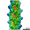


 PDBj
PDBj


 集合体
集合体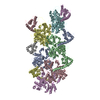

 試料調製
試料調製 電子顕微鏡撮影
 FIELD EMISSION GUN / 加速電圧: 300 kV / 照射モード: FLOOD BEAM
 解析
解析