- PDB-6ai7: Mandelate oxidase mutant-Y128F with the C4a-OH-FMN adduct -
+
Open data
ID or keywords:
Loading...
-
Basic information
Entry
Database: PDB / ID: 6ai7
Title
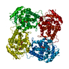
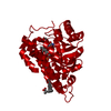
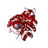
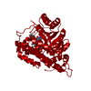
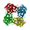
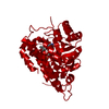
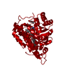
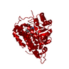
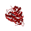
Mandelate oxidase mutant-Y128F with the C4a-OH-FMN adduct
Components
4-hydroxymandelate oxidase
Keywords
FLAVOPROTEIN / FMN-dependent oxidase
Function / homology
Function and homology information
4-hydroxymandelate oxidase / oxidoreductase activity, acting on the CH-OH group of donors, oxygen as acceptor / vancomycin biosynthetic process / FMN binding Similarity search - Function
Alpha-hydroxy acid dehydrogenase, FMN-dependent / FMN-dependent alpha-hydroxy acid dehydrogenase, active site / FMN hydroxy acid dehydrogenase domain / FMN-dependent alpha-hydroxy acid dehydrogenases active site. / FMN-dependent alpha-hydroxy acid dehydrogenase domain profile. / FMN-dependent dehydrogenase / FMN-dependent dehydrogenase / Aldolase class I / Aldolase-type TIM barrel / TIM Barrel ...Alpha-hydroxy acid dehydrogenase, FMN-dependent / FMN-dependent alpha-hydroxy acid dehydrogenase, active site / FMN hydroxy acid dehydrogenase domain / FMN-dependent alpha-hydroxy acid dehydrogenases active site. / FMN-dependent alpha-hydroxy acid dehydrogenase domain profile. / FMN-dependent dehydrogenase / FMN-dependent dehydrogenase / Aldolase class I / Aldolase-type TIM barrel / TIM Barrel / Alpha-Beta Barrel / Alpha Beta Similarity search - Domain/homology
Resolution: 2.07→30 Å / Cor.coef. Fo:Fc: 0.94 / Cor.coef. Fo:Fc free: 0.914 / SU B: 3.903 / SU ML: 0.107 / Cross valid method: THROUGHOUT / ESU R: 0.163 / ESU R Free: 0.157 / Details: HYDROGENS HAVE BEEN ADDED IN THE RIDING POSITIONS
Rfactor
Num. reflection
% reflection
Selection details
Rfree
0.24618
1561
5 %
RANDOM
Rwork
0.20475
-
-
-
obs
0.20681
29522
96.23 %
-
Solvent computation
Ion probe radii: 0.8 Å / Shrinkage radii: 0.8 Å / VDW probe radii: 1.2 Å
 Movie
Movie Controller


 Open data
 Basic information
 Components
 Keywords
 Function and homology information
 Amycolatopsis orientalis (bacteria)
 X-RAY DIFFRACTION /
  Authors
 Citation
 Structure visualization
 Downloads & links
 Other downloads


 PDBj
PDBj
 Assembly


 Mass: 474.359 Da / Num. of mol.: 1 / Source method: obtained synthetically / Formula: C17H23N4O10P
 Mass: 18.015 Da / Num. of mol.: 74 / Source method: isolated from a natural source / Formula: H2O
 Sample preparation
 / Beamline: BL15A1 / Wavelength: 1 Å
 Processing