[English] 日本語
Yorodumi
- PDB-6a39: The crystal structure of Mandelate oxidase Y128F with C4a-Malic a... -

+
Open data


ID or keywords:

Loading...

-
Basic information

Entry
Database: PDB / ID: 6a39
Title
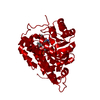
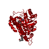
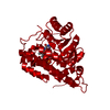
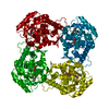
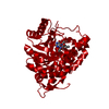
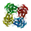
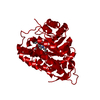
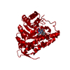
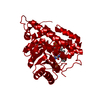
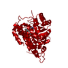
The crystal structure of Mandelate oxidase Y128F with C4a-Malic acid-monooxide-FMN adduct
Components4-hydroxymandelate oxidase
KeywordsFLAVOPROTEIN / FMN-dependent oxidase
Function / homology
Function and homology information


4-hydroxymandelate oxidase / oxidoreductase activity, acting on the CH-OH group of donors, oxygen as acceptor / vancomycin biosynthetic process / : / fatty acid alpha-oxidation / L-lactate dehydrogenase activity / peroxisome / FMN binding / plasma membrane
Similarity search - Function
Alpha-hydroxy acid dehydrogenase, FMN-dependent / FMN-dependent alpha-hydroxy acid dehydrogenase, active site / FMN hydroxy acid dehydrogenase domain / FMN-dependent alpha-hydroxy acid dehydrogenases active site. / FMN-dependent alpha-hydroxy acid dehydrogenase domain profile. / FMN-dependent dehydrogenase / FMN-dependent dehydrogenase / Aldolase-type TIM barrel
Similarity search - Domain/homology
Chem-B8C / 4-hydroxymandelate oxidase
Similarity search - Component
Biological speciesAmycolatopsis orientalis (bacteria)
MethodX-RAY DIFFRACTION / SYNCHROTRON / MOLECULAR REPLACEMENT / Resolution: 1.89 Å
AuthorsLi, T.L. / Lin, K.H.
CitationJournal: To Be Published
Title: The crystal structure of Mandelate oxidase mutant Y128F with C4a-Malic acid-monooxide-FMN adduct
Authors: Li, T.L. / Lin, K.H.
History
DepositionJun 15, 2018Deposition site: PDBJ / Processing site: PDBJ
Revision 1.0Jun 19, 2019Provider: repository / Type: Initial release
Revision 1.1Nov 22, 2023Group: Data collection / Database references / Refinement description
Category: chem_comp_atom / chem_comp_bond ...chem_comp_atom / chem_comp_bond / database_2 / pdbx_initial_refinement_model
Item: _database_2.pdbx_DOI / _database_2.pdbx_database_accession

-
Structure visualization

Structure viewerMolecule:
MolmilJmol/JSmol

Downloads & links

-
Assembly

Deposited unit
A: 4-hydroxymandelate oxidase
hetero molecules


Theoretical massNumber of molelcules
Total (without water)40,6342
Polymers40,0301
Non-polymers6041
Water4,756264
1
A: 4-hydroxymandelate oxidase
hetero molecules

A: 4-hydroxymandelate oxidase
hetero molecules

A: 4-hydroxymandelate oxidase
hetero molecules

A: 4-hydroxymandelate oxidase
hetero molecules


Theoretical massNumber of molelcules
Total (without water)162,5368
Polymers160,1184
Non-polymers2,4184
Water724
TypeNameSymmetry operationNumber
identity operation1_555x,y,z1
crystal symmetry operation2_555-x,-y,z1
crystal symmetry operation3_555-y,x,z1
crystal symmetry operation4_555y,-x,z1
Buried area9300 Å2
ΔGint-76 kcal/mol
Surface area44800 Å2
MethodPISA
Unit cell
Length a, b, c (Å)138.440, 138.440, 109.144
Angle α, β, γ (deg.)90.00, 90.00, 90.00
Int Tables number97
Space group name H-MI422

-
Components

#1: Protein 4-hydroxymandelate oxidase


Mass: 40029.562 Da / Num. of mol.: 1 / Mutation: Y128F
Source method: isolated from a genetically manipulated source
Source: (gene. exp.) Amycolatopsis orientalis (bacteria) / Gene: hmo / Production host: Escherichia coli (E. coli) / References: UniProt: O52792, 4-hydroxymandelate oxidase
#2: Chemical ChemComp-B8C / (~{E})-2-[[(4~{a}~{S})-7,8-dimethyl-2,4-bis(oxidanylidene)-10-[(2~{S},3~{S},4~{S})-2,3,4-tris(oxidanyl)-5-phosphonooxy-pentyl]-5~{H}-benzo[g]pteridin-4~{a}-yl]oxy]-3-oxidanyl-but-2-enedioic acid


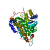
Mass: 604.415 Da / Num. of mol.: 1 / Source method: isolated from a natural source / Formula: C21H25N4O15P
#3: Water ChemComp-HOH / water


Mass: 18.015 Da / Num. of mol.: 264 / Source method: isolated from a natural source / Formula: H2O

-
Experimental details

-
Experiment

ExperimentMethod: X-RAY DIFFRACTION / Number of used crystals: 1

-
Sample preparation

CrystalDensity Matthews: 3.45 Å3/Da / Density % sol: 64.38 %
Crystal growTemperature: 293 K / Method: vapor diffusion, hanging drop / pH: 7 / Details: 35% Tascimate, 0.1M Bis-Tris propane pH 7.0

-
Data collection

DiffractionMean temperature: 100 K / Serial crystal experiment: N
Diffraction sourceSource: SYNCHROTRON / Site: NSRRC / Beamline: BL15A1 / Wavelength: 1 Å
DetectorType: RAYONIX MX300HE / Detector: CCD / Date: Apr 19, 2018
RadiationProtocol: SINGLE WAVELENGTH / Monochromatic (M) / Laue (L): M / Scattering type: x-ray
Radiation wavelengthWavelength: 1 Å / Relative weight: 1
ReflectionResolution: 1.894→30 Å / Num. obs: 40612 / % possible obs: 98.9 % / Redundancy: 9.7 % / Rmerge(I) obs: 0.039 / Rpim(I) all: 0.019 / Net I/σ(I): 2.62
Reflection shellResolution: 1.894→1.97 Å / Rmerge(I) obs: 0.745 / Num. unique obs: 4154 / CC1/2: 0.873 / Rpim(I) all: 0.275

-
Processing

Software
NameVersionClassification
REFMAC5.8.0238refinement
HKL-2000data reduction
HKL-2000data scaling
PHENIXphasing
RefinementMethod to determine structure: MOLECULAR REPLACEMENT
Starting model: 3SGZ
Resolution: 1.89→29.24 Å / Cor.coef. Fo:Fc: 0.95 / Cor.coef. Fo:Fc free: 0.92 / SU B: 3.021 / SU ML: 0.087 / Cross valid method: THROUGHOUT / ESU R: 0.118 / ESU R Free: 0.123 / Stereochemistry target values: MAXIMUM LIKELIHOOD
Details: SF FILE CONTAINS FRIEDEL PAIRS UNDER I/F_MINUS AND I/F_PLUS COLUMNS. HYDROGENS HAVE BEEN ADDED IN THE RIDING POSITIONS
RfactorNum. reflection% reflectionSelection details
Rfree0.235 2113 5.2 %RANDOM
Rwork0.18951 ---
obs0.19184 38499 96.05 %-
Solvent computationIon probe radii: 0.8 Å / Shrinkage radii: 0.8 Å / VDW probe radii: 1.2 Å / Solvent model: MASK
Displacement parametersBiso mean: 27.31 Å2
Baniso -1Baniso -2Baniso -3
1--0.09 Å2-0 Å2-0 Å2
2---0.09 Å2-0 Å2
3---0.19 Å2
Refinement stepCycle: 1 / Resolution: 1.89→29.24 Å
ProteinNucleic acidLigandSolventTotal
Num. atoms2458 0 42 264 2764
Refine LS restraints
Refine-IDTypeDev idealDev ideal targetNumber
X-RAY DIFFRACTIONr_bond_refined_d0.0240.0132547
X-RAY DIFFRACTIONr_bond_other_d0.0020.0172448
X-RAY DIFFRACTIONr_angle_refined_deg2.4831.6533467
X-RAY DIFFRACTIONr_angle_other_deg1.6641.5865601
X-RAY DIFFRACTIONr_dihedral_angle_1_deg7.5125330
X-RAY DIFFRACTIONr_dihedral_angle_2_deg26.64719.635137
X-RAY DIFFRACTIONr_dihedral_angle_3_deg16.31315396
X-RAY DIFFRACTIONr_dihedral_angle_4_deg16.5871530
X-RAY DIFFRACTIONr_chiral_restr0.3280.2326
X-RAY DIFFRACTIONr_gen_planes_refined0.0170.022918
X-RAY DIFFRACTIONr_gen_planes_other0.0010.02574
X-RAY DIFFRACTIONr_nbd_refined
X-RAY DIFFRACTIONr_nbd_other
X-RAY DIFFRACTIONr_nbtor_refined
X-RAY DIFFRACTIONr_nbtor_other
X-RAY DIFFRACTIONr_xyhbond_nbd_refined
X-RAY DIFFRACTIONr_xyhbond_nbd_other
X-RAY DIFFRACTIONr_metal_ion_refined
X-RAY DIFFRACTIONr_metal_ion_other
X-RAY DIFFRACTIONr_symmetry_vdw_refined
X-RAY DIFFRACTIONr_symmetry_vdw_other
X-RAY DIFFRACTIONr_symmetry_hbond_refined
X-RAY DIFFRACTIONr_symmetry_hbond_other
X-RAY DIFFRACTIONr_symmetry_metal_ion_refined
X-RAY DIFFRACTIONr_symmetry_metal_ion_other
X-RAY DIFFRACTIONr_mcbond_it4.2492.5041320
X-RAY DIFFRACTIONr_mcbond_other4.252.5011319
X-RAY DIFFRACTIONr_mcangle_it5.5073.7221647
X-RAY DIFFRACTIONr_mcangle_other5.5073.7251648
X-RAY DIFFRACTIONr_scbond_it5.6453.2331226
X-RAY DIFFRACTIONr_scbond_other5.6463.2331225
X-RAY DIFFRACTIONr_scangle_it
X-RAY DIFFRACTIONr_scangle_other7.9224.611820
X-RAY DIFFRACTIONr_long_range_B_refined10.26732.8443093
X-RAY DIFFRACTIONr_long_range_B_other10.26532.8413094
X-RAY DIFFRACTIONr_rigid_bond_restr
X-RAY DIFFRACTIONr_sphericity_free
X-RAY DIFFRACTIONr_sphericity_bonded
LS refinement shellResolution: 1.894→1.943 Å / Total num. of bins used: 20
RfactorNum. reflection% reflection
Rfree0.299 130 -
Rwork0.279 2266 -
obs--77.84 %

+
About Yorodumi

-
News

-
Feb 9, 2022. New format data for meta-information of EMDB entries

New format data for meta-information of EMDB entries

  • Version 3 of the EMDB header file is now the official format.
  • The previous official version 1.9 will be removed from the archive.

Related info.:EMDB header

External links:wwPDB to switch to version 3 of the EMDB data model

-
Aug 12, 2020. Covid-19 info

Covid-19 info

URL: https://pdbjlvh1.pdbj.org/emnavi/covid19.php

New page: Covid-19 featured information page in EM Navigator.

Related info.:Covid-19 info / Mar 5, 2020. Novel coronavirus structure data

+
Mar 5, 2020. Novel coronavirus structure data

Novel coronavirus structure data

Related info.:Yorodumi Speices / Aug 12, 2020. Covid-19 info

External links:COVID-19 featured content - PDBj / Molecule of the Month (242):Coronavirus Proteases

+
Jan 31, 2019. EMDB accession codes are about to change! (news from PDBe EMDB page)

EMDB accession codes are about to change! (news from PDBe EMDB page)

  • The allocation of 4 digits for EMDB accession codes will soon come to an end. Whilst these codes will remain in use, new EMDB accession codes will include an additional digit and will expand incrementally as the available range of codes is exhausted. The current 4-digit format prefixed with “EMD-” (i.e. EMD-XXXX) will advance to a 5-digit format (i.e. EMD-XXXXX), and so on. It is currently estimated that the 4-digit codes will be depleted around Spring 2019, at which point the 5-digit format will come into force.
  • The EM Navigator/Yorodumi systems omit the EMD- prefix.

Related info.:Q: What is EMD? / ID/Accession-code notation in Yorodumi/EM Navigator

External links:EMDB Accession Codes are Changing Soon! / Contact to PDBj

+
Jul 12, 2017. Major update of PDB

Major update of PDB

  • wwPDB released updated PDB data conforming to the new PDBx/mmCIF dictionary.
  • This is a major update changing the version number from 4 to 5, and with Remediation, in which all the entries are updated.
  • In this update, many items about electron microscopy experimental information are reorganized (e.g. em_software).
  • Now, EM Navigator and Yorodumi are based on the updated data.

External links:wwPDB Remediation / Enriched Model Files Conforming to OneDep Data Standards Now Available in the PDB FTP Archive

-
Yorodumi

Thousand views of thousand structures

  • Yorodumi is a browser for structure data from EMDB, PDB, SASBDB, etc.
  • This page is also the successor to EM Navigator detail page, and also detail information page/front-end page for Omokage search.
  • The word "yorodu" (or yorozu) is an old Japanese word meaning "ten thousand". "mi" (miru) is to see.

Related info.:EMDB / PDB / SASBDB / Comparison of 3 databanks / Yorodumi Search / Aug 31, 2016. New EM Navigator & Yorodumi / Yorodumi Papers / Jmol/JSmol / Function and homology information / Changes in new EM Navigator and Yorodumi

Read more