 Movie
Movie Controller

[English] 日本語
 Yorodumi
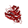
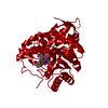
- PDB-6a0g: The crystal structure of Mandelate oxidase mutant Y128F with b-Ph... -
+ Open data

- Basic information
| Entry | Database: PDB / ID: 6a0g | ||||||
|---|---|---|---|---|---|---|---|
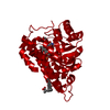
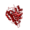
| Title | The crystal structure of Mandelate oxidase mutant Y128F with b-Phenyllactate | ||||||
 Components Components | 4-hydroxymandelate oxidase | ||||||
 Keywords Keywords | FLAVOPROTEIN / FMN-dependent oxidase | ||||||
| Function / homology |  Function and homology information Function and homology information4-hydroxymandelate oxidase / oxidoreductase activity, acting on the CH-OH group of donors, oxygen as acceptor / vancomycin biosynthetic process / FMN binding Similarity search - Function | ||||||
| Biological species |  Amycolatopsis orientalis (bacteria) Amycolatopsis orientalis (bacteria) | ||||||
| Method |  X-RAY DIFFRACTION / SYNCHROTRON / MOLECULAR REPLACEMENT / Resolution: 1.8 Å X-RAY DIFFRACTION / SYNCHROTRON / MOLECULAR REPLACEMENT / Resolution: 1.8 Å | ||||||
 Authors Authors | Li, T.L. / Lin, K.H. | ||||||
 Citation Citation | Journal: Acta Crystallogr D Struct Biol / Year: 2019 Title: Biochemical and structural explorations of alpha-hydroxyacid oxidases reveal a four-electron oxidative decarboxylation reaction. Authors: Yeh, H.W. / Lin, K.H. / Lyu, S.Y. / Li, Y.S. / Huang, C.M. / Wang, Y.L. / Shih, H.W. / Hsu, N.S. / Wu, C.J. / Li, T.L. | ||||||
| History |
|
- Structure visualization
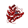
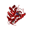
| Structure viewer | Molecule: MolmilJmol/JSmol |
|---|
- Downloads & links
-Download
| PDBx/mmCIF format | 6a0g.cif.gz | 89.6 KB | Display | PDBx/mmCIF format |
|---|---|---|---|---|
| PDB format | pdb6a0g.ent.gz | 64.9 KB | Display | PDB format |
| PDBx/mmJSON format | 6a0g.json.gz | Tree view | PDBx/mmJSON format | |
| Others |  Other downloads Other downloads |
-Validation report
| Arichive directory | https://data.pdbj.org/pub/pdb/validation_reports/a0/6a0gftp://data.pdbj.org/pub/pdb/validation_reports/a0/6a0g | HTTPS FTP |
|---|
-Related structure data
| Related structure data |  5zzqC 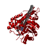 5zzsC  5zzxC  5zzyC  6a00C  6a0dC  6a0mC  6a0oC 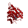 6a0yC  6a11C  6a1aC  3sgzS S: Starting model for refinement C: citing same article ( |
|---|---|
| Similar structure data |


-Links
 PDBj
PDBj
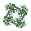
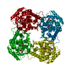
- Assembly
| Deposited unit | 
| ||||||||
|---|---|---|---|---|---|---|---|---|---|
| 1 | 
| ||||||||
| Unit cell |
|
-Components
| #1: Protein | Mass: 40029.562 Da / Num. of mol.: 1 / Mutation: Y128F Source method: isolated from a genetically manipulated source Source: (gene. exp.) Amycolatopsis orientalis (bacteria) / Gene: hmo / Production host: | ||
|---|---|---|---|
| #2: Chemical | ChemComp-FMN /   Mass: 456.344 Da / Num. of mol.: 1 / Source method: obtained synthetically / Formula: C17H21N4O9P Mass: 456.344 Da / Num. of mol.: 1 / Source method: obtained synthetically / Formula: C17H21N4O9P | ||
| #3: Chemical | 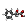  Type: L-peptide linking / Mass: 166.174 Da / Num. of mol.: 2 / Source method: obtained synthetically / Formula: C9H10O3 Type: L-peptide linking / Mass: 166.174 Da / Num. of mol.: 2 / Source method: obtained synthetically / Formula: C9H10O3#4: Water | ChemComp-HOH / |  Mass: 18.015 Da / Num. of mol.: 287 / Source method: isolated from a natural source / Formula: H2O Mass: 18.015 Da / Num. of mol.: 287 / Source method: isolated from a natural source / Formula: H2O |
-Experimental details
-Experiment
| Experiment | Method: X-RAY DIFFRACTION / Number of used crystals: 1 |
|---|
- Sample preparation
| Crystal | Density Matthews: 3.49 Å3/Da / Density % sol: 64.75 % |
|---|---|
| Crystal grow | Temperature: 293 K / Method: vapor diffusion, hanging drop / pH: 7 / Details: 35% Tascimate, 0.1M Bis-Tris propane pH 7.0 |
-Data collection
| Diffraction | Mean temperature: 100 K |
|---|---|
| Diffraction source | Source: SYNCHROTRON / Site: SPring-8  / Beamline: BL44XU / Wavelength: 1 Å / Beamline: BL44XU / Wavelength: 1 Å |
| Detector | Type: Bruker DIP-6040 / Detector: CCD / Date: Apr 23, 2016 |
| Radiation | Protocol: SINGLE WAVELENGTH / Monochromatic (M) / Laue (L): M / Scattering type: x-ray |
| Radiation wavelength | Wavelength: 1 Å / Relative weight: 1 |
| Reflection | Resolution: 1.8→30 Å / Num. obs: 49996 / % possible obs: 99.9 % / Redundancy: 11.1 % / Rmerge(I) obs: 0.048 / Rpim(I) all: 0.023 / Χ2: 0.947 / Net I/σ(I): 34.5 |
| Reflection shell | Resolution: 1.8→1.86 Å / Redundancy: 10.7 % / Rmerge(I) obs: 0.8 / Mean I/σ(I) obs: 2.7 / Num. unique obs: 4935 / CC1/2: 0.852 / Rpim(I) all: 0.32 / Χ2: 0.837 / % possible all: 100 |
- Processing
| Software |
| ||||||||||||||||||||||||||||||||||||||||||||||||||||||||||||||||||||||||||||||||||||||||||||||||||||||||||||||||||||||||||||||||||||||||||||||||||||||||||||||||||||||||||||||||||||||
|---|---|---|---|---|---|---|---|---|---|---|---|---|---|---|---|---|---|---|---|---|---|---|---|---|---|---|---|---|---|---|---|---|---|---|---|---|---|---|---|---|---|---|---|---|---|---|---|---|---|---|---|---|---|---|---|---|---|---|---|---|---|---|---|---|---|---|---|---|---|---|---|---|---|---|---|---|---|---|---|---|---|---|---|---|---|---|---|---|---|---|---|---|---|---|---|---|---|---|---|---|---|---|---|---|---|---|---|---|---|---|---|---|---|---|---|---|---|---|---|---|---|---|---|---|---|---|---|---|---|---|---|---|---|---|---|---|---|---|---|---|---|---|---|---|---|---|---|---|---|---|---|---|---|---|---|---|---|---|---|---|---|---|---|---|---|---|---|---|---|---|---|---|---|---|---|---|---|---|---|---|---|---|---|
| Refinement | Method to determine structure: MOLECULAR REPLACEMENT Starting model: 3SGZ Resolution: 1.8→30 Å / Cor.coef. Fo:Fc: 0.967 / Cor.coef. Fo:Fc free: 0.959 / SU B: 1.883 / SU ML: 0.057 / Cross valid method: THROUGHOUT / ESU R: 0.084 / ESU R Free: 0.083 / Details: HYDROGENS HAVE BEEN ADDED IN THE RIDING POSITIONS
| ||||||||||||||||||||||||||||||||||||||||||||||||||||||||||||||||||||||||||||||||||||||||||||||||||||||||||||||||||||||||||||||||||||||||||||||||||||||||||||||||||||||||||||||||||||||
| Solvent computation | Ion probe radii: 0.8 Å / Shrinkage radii: 0.8 Å / VDW probe radii: 1.2 Å | ||||||||||||||||||||||||||||||||||||||||||||||||||||||||||||||||||||||||||||||||||||||||||||||||||||||||||||||||||||||||||||||||||||||||||||||||||||||||||||||||||||||||||||||||||||||
| Displacement parameters | Biso mean: 25.616 Å2
| ||||||||||||||||||||||||||||||||||||||||||||||||||||||||||||||||||||||||||||||||||||||||||||||||||||||||||||||||||||||||||||||||||||||||||||||||||||||||||||||||||||||||||||||||||||||
| Refinement step | Cycle: 1 / Resolution: 1.8→30 Å
| ||||||||||||||||||||||||||||||||||||||||||||||||||||||||||||||||||||||||||||||||||||||||||||||||||||||||||||||||||||||||||||||||||||||||||||||||||||||||||||||||||||||||||||||||||||||
| Refine LS restraints |
|