[English] 日本語
 Yorodumi
- PDB-6adr: Anthrax Toxin Receptor 1-bound the Seneca Valley Virus in neutral... -
+ Open data
- Basic information
| Entry | Database: PDB / ID: 6adr | ||||||
|---|---|---|---|---|---|---|---|
| Title | Anthrax Toxin Receptor 1-bound the Seneca Valley Virus in neutral conditions | ||||||
 Components Components |
| ||||||
 Keywords Keywords | VIRUS / Seneca Valley virus | ||||||
| Function / homology |  Function and homology information Function and homology informationreproductive process / filopodium membrane / negative regulation of extracellular matrix assembly / blood vessel development / lamellipodium membrane / Uptake and function of anthrax toxins / collagen binding / substrate adhesion-dependent cell spreading / actin filament binding / transmembrane signaling receptor activity ...reproductive process / filopodium membrane / negative regulation of extracellular matrix assembly / blood vessel development / lamellipodium membrane / Uptake and function of anthrax toxins / collagen binding / substrate adhesion-dependent cell spreading / actin filament binding / transmembrane signaling receptor activity / actin cytoskeleton organization / endosome membrane / external side of plasma membrane / cell surface / metal ion binding / plasma membrane Similarity search - Function | ||||||
| Biological species |  Homo sapiens (human) Homo sapiens (human) Seneca valley virus Seneca valley virus | ||||||
| Method | ELECTRON MICROSCOPY / single particle reconstruction / cryo EM / Resolution: 3.38 Å | ||||||
 Authors Authors | Lou, Z.Y. / Cao, L. | ||||||
| Funding support |  China, 1items China, 1items
| ||||||
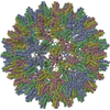
 Citation Citation |  Journal: Proc Natl Acad Sci U S A / Year: 2018 Journal: Proc Natl Acad Sci U S A / Year: 2018Title: Seneca Valley virus attachment and uncoating mediated by its receptor anthrax toxin receptor 1. Authors: Lin Cao / Ran Zhang / Tingting Liu / Zixian Sun / Mingxu Hu / Yuna Sun / Lingpeng Cheng / Yu Guo / Sheng Fu / Junjie Hu / Xiangmin Li / Chengqi Yu / Hanyang Wang / Huanchun Chen / Xueming Li ...Authors: Lin Cao / Ran Zhang / Tingting Liu / Zixian Sun / Mingxu Hu / Yuna Sun / Lingpeng Cheng / Yu Guo / Sheng Fu / Junjie Hu / Xiangmin Li / Chengqi Yu / Hanyang Wang / Huanchun Chen / Xueming Li / Elizabeth E Fry / David I Stuart / Ping Qian / Zhiyong Lou / Zihe Rao /   Abstract: Seneca Valley virus (SVV) is an oncolytic picornavirus with selective tropism for neuroendocrine cancers. SVV mediates cell entry by attachment to the receptor anthrax toxin receptor 1 (ANTXR1). Here ...Seneca Valley virus (SVV) is an oncolytic picornavirus with selective tropism for neuroendocrine cancers. SVV mediates cell entry by attachment to the receptor anthrax toxin receptor 1 (ANTXR1). Here we determine atomic structures of mature SVV particles alone and in complex with ANTXR1 in both neutral and acidic conditions, as well as empty "spent" particles in complex with ANTXR1 in acidic conditions by cryoelectron microscopy. SVV engages ANTXR1 mainly by the VP2 DF and VP1 CD loops, leading to structural changes in the VP1 GH loop and VP3 GH loop, which attenuate interprotomer interactions and destabilize the capsid assembly. Despite lying on the edge of the attachment site, VP2 D146 interacts with the metal ion in ANTXR1 and is required for cell entry. Though the individual substitution of most interacting residues abolishes receptor binding and virus propagation, a serine-to-alanine mutation at VP2 S177 significantly increases SVV proliferation. Acidification of the SVV-ANTXR1 complex results in a major reconfiguration of the pentameric capsid assemblies, which rotate ∼20° around the icosahedral fivefold axes to form a previously uncharacterized spent particle resembling a potential uncoating intermediate with remarkable perforations at both two- and threefold axes. These structures provide high-resolution snapshots of SVV entry, highlighting opportunities for anticancer therapeutic optimization. | ||||||
| History |
|
- Structure visualization
| Movie |
 Movie viewer Movie viewer |
|---|---|
| Structure viewer | Molecule:  Molmil Molmil Jmol/JSmol Jmol/JSmol |
- Downloads & links
- Download
| PDBx/mmCIF format |  6adr.cif.gz 6adr.cif.gz | 187.8 KB | Display |  PDBx/mmCIF format PDBx/mmCIF format |
|---|---|---|---|---|
| PDB format |  pdb6adr.ent.gz pdb6adr.ent.gz | 146.1 KB | Display |  PDB format PDB format |
| PDBx/mmJSON format |  6adr.json.gz 6adr.json.gz | Tree view |  PDBx/mmJSON format PDBx/mmJSON format | |
| Others |  Other downloads Other downloads |
-Validation report
| Summary document |  6adr_validation.pdf.gz 6adr_validation.pdf.gz | 997.5 KB | Display |  wwPDB validaton report wwPDB validaton report |
|---|---|---|---|---|
| Full document |  6adr_full_validation.pdf.gz 6adr_full_validation.pdf.gz | 1008.5 KB | Display | |
| Data in XML |  6adr_validation.xml.gz 6adr_validation.xml.gz | 33.7 KB | Display | |
| Data in CIF |  6adr_validation.cif.gz 6adr_validation.cif.gz | 50.4 KB | Display | |
| Arichive directory |  https://data.pdbj.org/pub/pdb/validation_reports/ad/6adr https://data.pdbj.org/pub/pdb/validation_reports/ad/6adr ftp://data.pdbj.org/pub/pdb/validation_reports/ad/6adr ftp://data.pdbj.org/pub/pdb/validation_reports/ad/6adr | HTTPS FTP |
-Related structure data
| Related structure data |  9611MC  9607C  9608C  9612C  9613C  6adlC  6admC  6adsC  6adtC M: map data used to model this data C: citing same article ( |
|---|---|
| Similar structure data |
- Links
Links
- Assembly
| Deposited unit | 
|
|---|---|
| 1 | x 60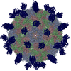
|
| 2 |
|
| 3 | x 5
|
| 4 | x 6
|
| 5 | 
|
| Symmetry | Point symmetry: (Schoenflies symbol: I (icosahedral)) |
- Components
-Protein , 5 types, 5 molecules ACBDR
| #1: Protein | Mass: 28494.023 Da / Num. of mol.: 1 / Source method: isolated from a natural source / Source: (natural)  Seneca valley virus Seneca valley virus |
|---|---|
| #2: Protein | Mass: 26393.938 Da / Num. of mol.: 1 / Source method: isolated from a natural source / Source: (natural)  Seneca valley virus Seneca valley virus |
| #3: Protein | Mass: 29843.289 Da / Num. of mol.: 1 / Source method: isolated from a natural source / Source: (natural)  Seneca valley virus Seneca valley virus |
| #4: Protein | Mass: 7393.875 Da / Num. of mol.: 1 / Source method: isolated from a natural source / Source: (natural)  Seneca valley virus Seneca valley virus |
| #5: Protein | Mass: 21316.020 Da / Num. of mol.: 1 / Fragment: UNP residues 38-220 / Mutation: C177A Source method: isolated from a genetically manipulated source Source: (gene. exp.)  Homo sapiens (human) / Gene: ANTXR1 / Production host: Homo sapiens (human) / Gene: ANTXR1 / Production host:  |
-Non-polymers , 2 types, 2 molecules 


| #6: Chemical | ChemComp-MG / |
|---|---|
| #7: Water | ChemComp-HOH / |
-Details
| Has protein modification | Y |
|---|
-Experimental details
-Experiment
| Experiment | Method: ELECTRON MICROSCOPY |
|---|---|
| EM experiment | Aggregation state: PARTICLE / 3D reconstruction method: single particle reconstruction |
- Sample preparation
| Component | Name: Seneca valley virus / Type: VIRUS / Entity ID: #1-#5 / Source: NATURAL |
|---|---|
| Source (natural) | Organism:  Seneca valley virus Seneca valley virus |
| Details of virus | Empty: YES / Enveloped: NO / Isolate: STRAIN / Type: VIRION |
| Buffer solution | pH: 6 |
| Specimen | Embedding applied: NO / Shadowing applied: NO / Staining applied: NO / Vitrification applied: YES |
| Vitrification | Cryogen name: ETHANE-PROPANE |
- Electron microscopy imaging
| Experimental equipment |  Model: Talos Arctica / Image courtesy: FEI Company |
|---|---|
| Microscopy | Model: FEI TECNAI ARCTICA |
| Electron gun | Electron source:  FIELD EMISSION GUN / Accelerating voltage: 200 kV / Illumination mode: FLOOD BEAM FIELD EMISSION GUN / Accelerating voltage: 200 kV / Illumination mode: FLOOD BEAM |
| Electron lens | Mode: BRIGHT FIELD |
| Image recording | Electron dose: 1.63 e/Å2 / Film or detector model: FEI FALCON II (4k x 4k) |
- Processing
| Software | Name: PHENIX / Version: 1.11.1_2575: / Classification: refinement | ||||||||||||||||||||||||
|---|---|---|---|---|---|---|---|---|---|---|---|---|---|---|---|---|---|---|---|---|---|---|---|---|---|
| CTF correction | Type: PHASE FLIPPING AND AMPLITUDE CORRECTION | ||||||||||||||||||||||||
| 3D reconstruction | Resolution: 3.38 Å / Resolution method: FSC 0.143 CUT-OFF / Num. of particles: 15460 / Symmetry type: POINT | ||||||||||||||||||||||||
| Atomic model building | Protocol: RIGID BODY FIT | ||||||||||||||||||||||||
| Refine LS restraints |
|
 Movie
Movie Controller






 PDBj
PDBj


