 ムービー
ムービー コントローラー


+ データを開く

- 基本情報
基本情報
| 登録情報 | データベース: PDB / ID: 6a70 | ||||||||||||
|---|---|---|---|---|---|---|---|---|---|---|---|---|---|
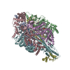
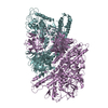
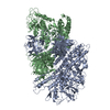
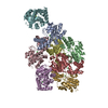
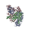
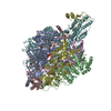
| タイトル | Structure of the human PKD1/PKD2 complex | ||||||||||||
 要素 要素 |
| ||||||||||||
 キーワード キーワード | MEMBRANE PROTEIN / Asymmetric complex / polycystic kidney disease | ||||||||||||
| 機能・相同性 |  機能・相同性情報 機能・相同性情報metanephric distal tubule morphogenesis / nitrogen cycle metabolic process / detection of nodal flow / metanephric smooth muscle tissue development / metanephric cortex development / metanephric cortical collecting duct development / metanephric distal tubule development / polycystin complex / mesonephric tubule development / mesonephric duct development ...metanephric distal tubule morphogenesis / nitrogen cycle metabolic process / detection of nodal flow / metanephric smooth muscle tissue development / metanephric cortex development / metanephric cortical collecting duct development / metanephric distal tubule development / polycystin complex / mesonephric tubule development / mesonephric duct development / metanephric part of ureteric bud development / renal tubule morphogenesis / determination of liver left/right asymmetry / HLH domain binding / lung epithelium development / metanephric ascending thin limb development / lymph vessel morphogenesis / metanephric mesenchyme development / metanephric S-shaped body morphogenesis / basal cortex / renal artery morphogenesis / mitocytosis / positive regulation of inositol 1,4,5-trisphosphate-sensitive calcium-release channel activity / metanephric proximal tubule development / calcium-induced calcium release activity / calcium-independent cell-matrix adhesion / Wnt receptor activity / migrasome / cilium organization / VxPx cargo-targeting to cilium / genitalia development / detection of mechanical stimulus / muscle alpha-actinin binding / regulation of calcium ion import / voltage-gated monoatomic ion channel activity / placenta blood vessel development / response to fluid shear stress / cellular response to hydrostatic pressure / Golgi-associated vesicle membrane / cation channel complex / cellular response to fluid shear stress / metanephric collecting duct development / outward rectifier potassium channel activity / actinin binding / cellular response to osmotic stress / non-motile cilium / digestive tract development / determination of left/right symmetry / voltage-gated monoatomic cation channel activity / inorganic cation transmembrane transport / aorta development / cartilage development / neural tube development / motile cilium / voltage-gated sodium channel activity / ciliary membrane / cartilage condensation / branching involved in ureteric bud morphogenesis / protein heterotetramerization / branching morphogenesis of an epithelial tube / skin development / negative regulation of G1/S transition of mitotic cell cycle / spinal cord development / establishment of cell polarity / cytoplasmic side of endoplasmic reticulum membrane / homophilic cell adhesion via plasma membrane adhesion molecules / heart looping / regulation of G1/S transition of mitotic cell cycle / centrosome duplication / voltage-gated potassium channel activity / cell surface receptor signaling pathway via JAK-STAT / anatomical structure morphogenesis / potassium channel activity / lateral plasma membrane / embryonic placenta development / voltage-gated calcium channel activity / regulation of cell adhesion / transcription regulator inhibitor activity / monoatomic cation channel activity / cytoskeletal protein binding / cellular response to cAMP / regulation of proteasomal protein catabolic process / release of sequestered calcium ion into cytosol / potassium ion transmembrane transport / calcium channel complex / sodium ion transmembrane transport / regulation of mitotic spindle organization / cellular response to calcium ion / cytoplasmic vesicle membrane / protein export from nucleus / liver development / basal plasma membrane / cell-matrix adhesion / kidney development / lumenal side of endoplasmic reticulum membrane / cellular response to reactive oxygen species / establishment of localization in cell / phosphoprotein binding / protein tetramerization / calcium ion transmembrane transport 類似検索 - 分子機能 | ||||||||||||
| 生物種 |  Homo sapiens (ヒト) Homo sapiens (ヒト) | ||||||||||||
| 手法 | 電子顕微鏡法 / 単粒子再構成法 / クライオ電子顕微鏡法 / 解像度: 3.6 Å | ||||||||||||
 データ登録者 データ登録者 | Su, Q. / Hu, F. / Ge, X. / Lei, J. / Yu, S. / Wang, T. / Zhou, Q. / Mei, C. / Shi, Y. | ||||||||||||
| 資金援助 |  中国, 3件 中国, 3件
| ||||||||||||
 引用 引用 | ジャーナル: Science / 年: 2018 タイトル: Structure of the human PKD1-PKD2 complex. 著者: Qiang Su / Feizhuo Hu / Xiaofei Ge / Jianlin Lei / Shengqiang Yu / Tingliang Wang / Qiang Zhou / Changlin Mei / Yigong Shi / 要旨: Mutations in two genes, and , account for most cases of autosomal dominant polycystic kidney disease, one of the most common monogenetic disorders. Here we report the 3.6-angstrom cryo-electron ...Mutations in two genes, and , account for most cases of autosomal dominant polycystic kidney disease, one of the most common monogenetic disorders. Here we report the 3.6-angstrom cryo-electron microscopy structure of truncated human PKD1-PKD2 complex assembled in a 1:3 ratio. PKD1 contains a voltage-gated ion channel (VGIC) fold that interacts with PKD2 to form the domain-swapped, yet noncanonical, transient receptor potential (TRP) channel architecture. The S6 helix in PKD1 is broken in the middle, with the extracellular half, S6a, resembling pore helix 1 in a typical TRP channel. Three positively charged, cavity-facing residues on S6b may block cation permeation. In addition to the VGIC, a five-transmembrane helix domain and a cytosolic PLAT domain were resolved in PKD1. The PKD1-PKD2 complex structure establishes a framework for dissecting the function and disease mechanisms of the PKD proteins. | ||||||||||||
| 履歴 |
|
- 構造の表示
構造の表示
| ムービー |
ムービービューア |
|---|---|
| 構造ビューア | 分子: MolmilJmol/JSmol |
- ダウンロードとリンク
-ダウンロード
| PDBx/mmCIF形式 | 6a70.cif.gz | 374.6 KB | 表示 | PDBx/mmCIF形式 |
|---|---|---|---|---|
| PDB形式 | pdb6a70.ent.gz | 281.1 KB | 表示 | PDB形式 |
| PDBx/mmJSON形式 | 6a70.json.gz | ツリー表示 | PDBx/mmJSON形式 | |
| その他 |  その他のダウンロード その他のダウンロード |
-検証レポート
| 文書・要旨 | 6a70_validation.pdf.gz | 1015.7 KB | 表示 | wwPDB検証レポート |
|---|---|---|---|---|
| 文書・詳細版 | 6a70_full_validation.pdf.gz | 1 MB | 表示 | |
| XML形式データ | 6a70_validation.xml.gz | 55.2 KB | 表示 | |
| CIF形式データ | 6a70_validation.cif.gz | 84.6 KB | 表示 | |
| アーカイブディレクトリ | https://data.pdbj.org/pub/pdb/validation_reports/a7/6a70ftp://data.pdbj.org/pub/pdb/validation_reports/a7/6a70 | HTTPS FTP |
-関連構造データ
| 関連構造データ |  6991MC  6992C M: このデータのモデリングに利用したマップデータ C: 同じ文献を引用 ( |
|---|---|
| 類似構造データ | |
| 電子顕微鏡画像生データ | EMPIAR-10262 (タイトル: Structure of the human PKD1-PKD2 complex / Data size: 209.8 Data #1: Autopicked particles of PKD1/PKD2 complex [picked particles - multiframe - unprocessed]) |


-リンク
 PDBj
PDBj


- 集合体
集合体
| 登録構造単位 | 
|
|---|---|
| 1 |
|
-要素
| #1: タンパク質 | 分子量: 66623.406 Da / 分子数: 3 / 由来タイプ: 組換発現 / 由来: (組換発現) Homo sapiens (ヒト) / 遺伝子: PKD2, TRPP2 / 細胞株 (発現宿主): HEK293F / 発現宿主: HOMO SAPIENS (ヒト) / 参照: UniProt: Q13563#2: タンパク質 | | 分子量: 127024.836 Da / 分子数: 1 / 由来タイプ: 組換発現 / 由来: (組換発現) Homo sapiens (ヒト) / 遺伝子: PKD1 / 細胞株 (発現宿主): HEK293F / 発現宿主: HOMO SAPIENS (ヒト) / 参照: UniProt: P98161 |
|---|
-実験情報
-実験
| 実験 | 手法: 電子顕微鏡法 |
|---|---|
| EM実験 | 試料の集合状態: PARTICLE / 3次元再構成法: 単粒子再構成法 |
- 試料調製
試料調製
| 構成要素 | 名称: the structure of an asymmetric complex / タイプ: COMPLEX 詳細: Samples were obtained by co-expression in 293F cells. A complex contains one PKD1 subunit and three PKD2 subunits. Entity ID: all / 由来: RECOMBINANT | ||||||||||||||||||||
|---|---|---|---|---|---|---|---|---|---|---|---|---|---|---|---|---|---|---|---|---|---|
| 分子量 | 値: 0.31 MDa / 実験値: NO | ||||||||||||||||||||
| 由来(天然) | 生物種: Homo sapiens (ヒト) | ||||||||||||||||||||
| 由来(組換発現) | 生物種: HOMO SAPIENS (ヒト) / 細胞: HEK293F | ||||||||||||||||||||
| 緩衝液 | pH: 7.5 | ||||||||||||||||||||
| 緩衝液成分 |
| ||||||||||||||||||||
| 試料 | 濃度: 10 mg/ml / 包埋: NO / シャドウイング: NO / 染色: NO / 凍結: YES / 詳細: This sample was monodisperse. | ||||||||||||||||||||
| 試料支持 | グリッドの材料: GOLD / グリッドのサイズ: 300 divisions/in. / グリッドのタイプ: Quantifoil R1.2/1.3 | ||||||||||||||||||||
| 急速凍結 | 装置: FEI VITROBOT MARK IV / 凍結剤: ETHANE / 湿度: 100 % / 凍結前の試料温度: 281 K |
- 電子顕微鏡撮影
| 実験機器 |  モデル: Titan Krios / 画像提供: FEI Company |
|---|---|
| 顕微鏡 | モデル: FEI TITAN KRIOS |
| 電子銃 | 電子線源:  FIELD EMISSION GUN / 加速電圧: 300 kV / 照射モード: FLOOD BEAM FIELD EMISSION GUN / 加速電圧: 300 kV / 照射モード: FLOOD BEAM |
| 電子レンズ | モード: DARK FIELD |
| 撮影 | 電子線照射量: 50 e/Å2 フィルム・検出器のモデル: GATAN K2 SUMMIT (4k x 4k) |
- 解析
解析
| ソフトウェア | 名称: PHENIX / バージョン: 1.13_2998: / 分類: 精密化 |
|---|---|
| CTF補正 | タイプ: PHASE FLIPPING AND AMPLITUDE CORRECTION |
| 3次元再構成 | 解像度: 3.6 Å / 解像度の算出法: FSC 0.143 CUT-OFF / 粒子像の数: 27296 / 対称性のタイプ: POINT |
