Movie
Movie Controller


+ Open data

- Basic information
| Entry | Database: EMDB / ID: EMD-6991 | ||||||||||||
|---|---|---|---|---|---|---|---|---|---|---|---|---|---|
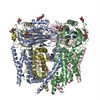
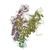
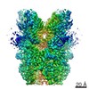
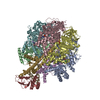
| Title | Structure of the human PKD1/PKD2 complex | ||||||||||||
 Map data Map data | None | ||||||||||||
 Sample Sample |
| ||||||||||||
 Keywords Keywords | Asymmetric complex / polycystic kidney disease / MEMBRANE PROTEIN | ||||||||||||
| Function / homology |  Function and homology information Function and homology informationmetanephric distal tubule morphogenesis / lung epithelium development / nitrogen cycle metabolic process / detection of nodal flow / metanephric smooth muscle tissue development / metanephric cortex development / metanephric cortical collecting duct development / metanephric distal tubule development / polycystin complex / mesonephric tubule development ...metanephric distal tubule morphogenesis / lung epithelium development / nitrogen cycle metabolic process / detection of nodal flow / metanephric smooth muscle tissue development / metanephric cortex development / metanephric cortical collecting duct development / metanephric distal tubule development / polycystin complex / mesonephric tubule development / mesonephric duct development / metanephric part of ureteric bud development / renal tubule morphogenesis / determination of liver left/right asymmetry / metanephric ascending thin limb development / lymph vessel morphogenesis / metanephric mesenchyme development / metanephric S-shaped body morphogenesis / basal cortex / mitocytosis / renal artery morphogenesis / placenta blood vessel development / metanephric proximal tubule development / HLH domain binding / calcium-independent cell-matrix adhesion / Wnt receptor activity / VxPx cargo-targeting to cilium / genitalia development / cilium organization / migrasome / muscle alpha-actinin binding / regulation of calcium ion import / cellular response to fluid shear stress / calcium-induced calcium release activity / detection of mechanical stimulus / voltage-gated monoatomic ion channel activity / Golgi-associated vesicle membrane / cellular response to hydrostatic pressure / response to fluid shear stress / metanephric collecting duct development / cation channel complex / digestive tract development / non-motile cilium / outward rectifier potassium channel activity / motile cilium / neural tube development / determination of left/right symmetry / cartilage development / cellular response to osmotic stress / actinin binding / aorta development / branching involved in ureteric bud morphogenesis / voltage-gated sodium channel activity / cartilage condensation / skin development / voltage-gated monoatomic cation channel activity / branching morphogenesis of an epithelial tube / ciliary membrane / negative regulation of G1/S transition of mitotic cell cycle / positive regulation of phospholipase C-activating G protein-coupled receptor signaling pathway / heart looping / protein heterotetramerization / establishment of cell polarity / spinal cord development / cytoplasmic side of endoplasmic reticulum membrane / homophilic cell-cell adhesion / centrosome duplication / embryonic placenta development / lateral plasma membrane / anatomical structure morphogenesis / voltage-gated potassium channel activity / potassium channel activity / regulation of cell adhesion / cell surface receptor signaling pathway via JAK-STAT / transcription regulator inhibitor activity / voltage-gated calcium channel activity / monoatomic cation channel activity / regulation of G1/S transition of mitotic cell cycle / cytoskeletal protein binding / release of sequestered calcium ion into cytosol / protein export from nucleus / calcium channel complex / potassium ion transmembrane transport / cellular response to calcium ion / regulation of proteasomal protein catabolic process / cytoplasmic vesicle membrane / basal plasma membrane / cellular response to cAMP / cell-matrix adhesion / liver development / kidney development / regulation of mitotic spindle organization / sodium ion transmembrane transport / lumenal side of endoplasmic reticulum membrane / protein tetramerization / cellular response to reactive oxygen species / phosphoprotein binding / Wnt signaling pathway / transmembrane transport / calcium channel activity Similarity search - Function | ||||||||||||
| Biological species |  Homo sapiens (human) Homo sapiens (human) | ||||||||||||
| Method | single particle reconstruction / cryo EM / Resolution: 3.6 Å | ||||||||||||
 Authors Authors | Su Q / Hu F / Ge X / Lei J / Yu S / Wang T / Zhou Q / Mei C / Shi Y | ||||||||||||
| Funding support |  China, 3 items China, 3 items
| ||||||||||||
 Citation Citation | Journal: Science / Year: 2018 Title: Structure of the human PKD1-PKD2 complex. Authors: Qiang Su / Feizhuo Hu / Xiaofei Ge / Jianlin Lei / Shengqiang Yu / Tingliang Wang / Qiang Zhou / Changlin Mei / Yigong Shi / Abstract: Mutations in two genes, and , account for most cases of autosomal dominant polycystic kidney disease, one of the most common monogenetic disorders. Here we report the 3.6-angstrom cryo-electron ...Mutations in two genes, and , account for most cases of autosomal dominant polycystic kidney disease, one of the most common monogenetic disorders. Here we report the 3.6-angstrom cryo-electron microscopy structure of truncated human PKD1-PKD2 complex assembled in a 1:3 ratio. PKD1 contains a voltage-gated ion channel (VGIC) fold that interacts with PKD2 to form the domain-swapped, yet noncanonical, transient receptor potential (TRP) channel architecture. The S6 helix in PKD1 is broken in the middle, with the extracellular half, S6a, resembling pore helix 1 in a typical TRP channel. Three positively charged, cavity-facing residues on S6b may block cation permeation. In addition to the VGIC, a five-transmembrane helix domain and a cytosolic PLAT domain were resolved in PKD1. The PKD1-PKD2 complex structure establishes a framework for dissecting the function and disease mechanisms of the PKD proteins. | ||||||||||||
| History |
|
- Structure visualization
| Movie |
Movie viewer |
|---|---|
| Structure viewer | EM map: SurfViewMolmilJmol/JSmol |
| Supplemental images |

- Downloads & links
-EMDB archive
| Map data | emd_6991.map.gz | 58.7 MB | EMDB map data format | |
|---|---|---|---|---|
| Header (meta data) | emd-6991-v30.xmlemd-6991.xml | 14.5 KB 14.5 KB | Display Display | EMDB header |
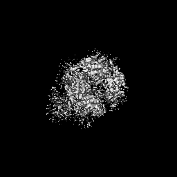
| Images | 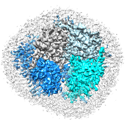 emd_6991.png emd_6991.png | 238.9 KB | ||
| Filedesc metadata | emd-6991.cif.gz | 6.4 KB | ||
| Archive directory |  http://ftp.pdbj.org/pub/emdb/structures/EMD-6991ftp://ftp.pdbj.org/pub/emdb/structures/EMD-6991 http://ftp.pdbj.org/pub/emdb/structures/EMD-6991ftp://ftp.pdbj.org/pub/emdb/structures/EMD-6991 | HTTPS FTP |
-Related structure data
| Related structure data |  6a70MC  6992C C: citing same article ( M: atomic model generated by this map |
|---|---|
| Similar structure data | |
| EM raw data | EMPIAR-10262 (Title: Structure of the human PKD1-PKD2 complex / Data size: 209.8 Data #1: Autopicked particles of PKD1/PKD2 complex [picked particles - multiframe - unprocessed]) |


-Links
| EMDB pages | EMDB (EBI/PDBe) / EMDataResource |
|---|---|
| Related items in Molecule of the Month |


-Map
| File | Download / File: emd_6991.map.gz / Format: CCP4 / Size: 64 MB / Type: IMAGE STORED AS FLOATING POINT NUMBER (4 BYTES) | ||||||||||||||||||||||||||||||||||||||||||||||||||||||||||||
|---|---|---|---|---|---|---|---|---|---|---|---|---|---|---|---|---|---|---|---|---|---|---|---|---|---|---|---|---|---|---|---|---|---|---|---|---|---|---|---|---|---|---|---|---|---|---|---|---|---|---|---|---|---|---|---|---|---|---|---|---|---|
| Annotation | None | ||||||||||||||||||||||||||||||||||||||||||||||||||||||||||||
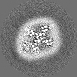
| Projections & slices | Image control
Images are generated by Spider. | ||||||||||||||||||||||||||||||||||||||||||||||||||||||||||||
| Voxel size | X=Y=Z: 1.091 Å | ||||||||||||||||||||||||||||||||||||||||||||||||||||||||||||
| Density |
| ||||||||||||||||||||||||||||||||||||||||||||||||||||||||||||
| Symmetry | Space group: 1 | ||||||||||||||||||||||||||||||||||||||||||||||||||||||||||||
| Details | EMDB XML:
CCP4 map header:
| ||||||||||||||||||||||||||||||||||||||||||||||||||||||||||||
 Z (Sec.)
 Y (Row.)
 X (Col.)


-Supplemental data
- Sample components
-Entire : the structure of an asymmetric complex
| Entire | Name: the structure of an asymmetric complex |
|---|---|
| Components |
|
-Supramolecule #1: the structure of an asymmetric complex
| Supramolecule | Name: the structure of an asymmetric complex / type: complex / ID: 1 / Parent: 0 / Macromolecule list: all Details: Samples were obtained by co-expression in 293F cells. A complex contains one PKD1 subunit and three PKD2 subunits. |
|---|---|
| Source (natural) | Organism: Homo sapiens (human) |
| Molecular weight | Theoretical: 310 KDa |
-Macromolecule #1: Polycystin-2
| Macromolecule | Name: Polycystin-2 / type: protein_or_peptide / ID: 1 / Number of copies: 3 / Enantiomer: LEVO |
|---|---|
| Source (natural) | Organism: Homo sapiens (human) |
| Molecular weight | Theoretical: 66.623406 KDa |
| Recombinant expression | Organism: HOMO SAPIENS (human) |
| Sequence | String: MGSAGWSHPQ FEKGGGSGGG SGGSAWSHPQ FEKGSAAAPR VAWAERLVRG LRGLWGTRLM EESSTNREKY LKSVLRELVT YLLFLIVLC ILTYGMMSSN VYYYTRMMSQ LFLDTPVSKT EKTNFKTLSS MEDFWKFTEG SLLDGLYWKM QPSNQTEADN R SFIFYENL ...String: MGSAGWSHPQ FEKGGGSGGG SGGSAWSHPQ FEKGSAAAPR VAWAERLVRG LRGLWGTRLM EESSTNREKY LKSVLRELVT YLLFLIVLC ILTYGMMSSN VYYYTRMMSQ LFLDTPVSKT EKTNFKTLSS MEDFWKFTEG SLLDGLYWKM QPSNQTEADN R SFIFYENL LLGVPRIRQL RVRNGSCSIP QDLRDEIKEC YDVYSVSSED RAPFGPRNGT AWIYTSEKDL NGSSHWGIIA TY SGAGYYL DLSRTREETA AQVASLKKNV WLDRGTRATF IDFSVYNANI NLFCVVRLLV EFPATGGVIP SWQFQPLKLI RYV TTFDFF LAACEIIFCF FIFYYVVEEI LEIRIHKLHY FRSFWNCLDV VIVVLSVVAI GINIYRTSNV EVLLQFLEDQ NTFP NFEHL AYWQIQFNNI AAVTVFFVWI KLFKFINFNR TMSQLSTTMS RCAKDLFGFA IMFFIIFLAY AQLAYLVFGT QVDDF STFQ ECIFTQFRII LGDINFAEIE EANRVLGPIY FTTFVFFMFF ILLNMFLAII NDTYSEVKSD LAQQKAEMEL SDLIRK GYH KALVKLKLKK NTVD UniProtKB: Polycystin-2 |
-Macromolecule #2: Polycystin-1
| Macromolecule | Name: Polycystin-1 / type: protein_or_peptide / ID: 2 / Number of copies: 1 / Enantiomer: LEVO |
|---|---|
| Source (natural) | Organism: Homo sapiens (human) |
| Molecular weight | Theoretical: 127.024836 KDa |
| Recombinant expression | Organism: HOMO SAPIENS (human) |
| Sequence | String: MGSAGDYKDH DGDYKDHDID YKDDDDKGSA AATAFGASLF VPPSHVRFVF PEPTADVNYI VMLTCAVCLV TYMVMAAILH KLDQLDASR GRAIPFCGQR GRFKYEILVK TGWGRGSGTT AHVGIMLYGV DSRSGHRHLD GDRAFHRNSL DIFRIATPHS L GSVWKIRV ...String: MGSAGDYKDH DGDYKDHDID YKDDDDKGSA AATAFGASLF VPPSHVRFVF PEPTADVNYI VMLTCAVCLV TYMVMAAILH KLDQLDASR GRAIPFCGQR GRFKYEILVK TGWGRGSGTT AHVGIMLYGV DSRSGHRHLD GDRAFHRNSL DIFRIATPHS L GSVWKIRV WHDNKGLSPA WFLQHVIVRD LQTARSAFFL VNDWLSVETE ANGGLVEKEV LAASDAALLR FRRLLVAELQ RG FFDKHIW LSIWDRPPRS RFTRIQRATC CVLLICLFLG ANAVWYGAVG DSAYSTGHVS RLSPLSVDTV AVGLVSSVVV YPV YLAILF LFRMSRSKVA GSPSPTPAGQ QVLDIDSCLD SSVLDSSFLT FSGLHAEQAF VGQMKSDLFL DDSKSLVCWP SGEG TLSWP DLLSDPSIVG SNLRQLARGQ AGHGLGPEED GFSLASPYSP AKSFSASDED LIQQVLAEGV SSPAPTQDTH METDL LSSL SSTPGEKTET LALQRLGELG PPSPGLNWEQ PQAARLSRTG LVEGLRKRLL PAWCASLAHG LSLLLVAVAV AVSGWV GAS FPPGVSVAWL LSSSASFLAS FLGWEPLKVL LEALYFSLVA KRLHPDEDDT LVESPAVTPV SARVPRVRPP HGFALFL AK EEARKVKRLH GMLRSLLVYM LFLLVTLLAS YGDASCHGHA YRLQSAIKQE LHSRAFLAIT RSEELWPWMA HVLLPYVH G NQSSPELGPP RLRQVRLQEA LYPDPPGPRV HTCSAAGGFS TSDYDVGWES PHNGSGTWAY SAPDLLGAWS WGSCAVYDS GGYVQELGLS LEESRDRLRF LQLHNWLDNR SRAVFLELTR YSPAVGLHAA VTLRLEFPAA GRALAALSVR PFALRRLSAG LSLPLLTSV CLLLFAVHFA VAEARTWHRE GRWRVLRLGA WARWLLVALT AATALVRLAQ LGAADRQWTR FVRGRPRRFT S FDQVAQLS SAARGLAASL LFLLLVKAAQ QLRFVRQWSV FGKTLCRALP ELLGVTLGLV VLGVAYAQLA ILLVSSCVDS LW SVAQALL VLCPGTGLST LCPAESWHLS PLLCVGLWAL RLWGALRLGA VILRWRYHAL RGELYRPAWE PQDYEMVELF LRR LRLWMG LSKVKEFRHK VRFEGMEPLP SRSSRGS UniProtKB: Polycystin-1 |
-Experimental details
-Structure determination
| Method | cryo EM |
|---|---|
 Processing Processing | single particle reconstruction |
| Aggregation state | particle |
-Sample preparation
| Concentration | 10 mg/mL | ||||||||||||
|---|---|---|---|---|---|---|---|---|---|---|---|---|---|
| Buffer | pH: 7.5 Component:
| ||||||||||||
| Grid | Model: Quantifoil R1.2/1.3 / Material: GOLD / Mesh: 300 / Support film - Material: CARBON / Pretreatment - Type: GLOW DISCHARGE / Pretreatment - Time: 30 sec. | ||||||||||||
| Vitrification | Cryogen name: ETHANE / Chamber humidity: 100 % / Chamber temperature: 281 K / Instrument: FEI VITROBOT MARK IV | ||||||||||||
| Details | This sample was monodisperse. |
- Electron microscopy
| Microscope | FEI TITAN KRIOS |
|---|---|
| Image recording | Film or detector model: GATAN K2 SUMMIT (4k x 4k) / Average electron dose: 50.0 e/Å2 |
| Electron beam | Acceleration voltage: 300 kV / Electron source:  FIELD EMISSION GUN FIELD EMISSION GUN |
| Electron optics | Illumination mode: FLOOD BEAM / Imaging mode: DARK FIELD |
| Experimental equipment |  Model: Titan Krios / Image courtesy: FEI Company |
-Image processing
| Startup model | Type of model: PDB ENTRY PDB model - PDB ID: |
|---|---|
| Final reconstruction | Resolution.type: BY AUTHOR / Resolution: 3.6 Å / Resolution method: FSC 0.143 CUT-OFF / Number images used: 27296 |
| Initial angle assignment | Type: ANGULAR RECONSTITUTION |
| Final angle assignment | Type: ANGULAR RECONSTITUTION |