 Movie
Movie Controller

[English] 日本語
 Yorodumi
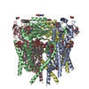
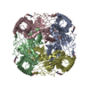
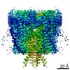
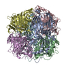
- PDB-5t4d: Cryo-EM structure of Polycystic Kidney Disease protein 2 (PKD2), ... -
+ Open data

- Basic information
| Entry | Database: PDB / ID: 5t4d | ||||||
|---|---|---|---|---|---|---|---|
| Title | Cryo-EM structure of Polycystic Kidney Disease protein 2 (PKD2), residues 198-703 | ||||||
 Components Components | hPKD:198-703, Polycystin-2 | ||||||
 Keywords Keywords | METAL TRANSPORT / TRP channel / PKD2 / nanodisc / TRPP | ||||||
| Function / homology |  Function and homology information Function and homology informationdetection of nodal flow / metanephric smooth muscle tissue development / metanephric cortex development / metanephric cortical collecting duct development / metanephric distal tubule development / polycystin complex / mesonephric tubule development / mesonephric duct development / metanephric part of ureteric bud development / renal tubule morphogenesis ...detection of nodal flow / metanephric smooth muscle tissue development / metanephric cortex development / metanephric cortical collecting duct development / metanephric distal tubule development / polycystin complex / mesonephric tubule development / mesonephric duct development / metanephric part of ureteric bud development / renal tubule morphogenesis / determination of liver left/right asymmetry / metanephric ascending thin limb development / metanephric mesenchyme development / metanephric S-shaped body morphogenesis / basal cortex / renal artery morphogenesis / placenta blood vessel development / HLH domain binding / VxPx cargo-targeting to cilium / cilium organization / migrasome / muscle alpha-actinin binding / regulation of calcium ion import / cellular response to fluid shear stress / calcium-induced calcium release activity / detection of mechanical stimulus / voltage-gated monoatomic ion channel activity / cellular response to hydrostatic pressure / cation channel complex / non-motile cilium / outward rectifier potassium channel activity / motile cilium / neural tube development / determination of left/right symmetry / cellular response to osmotic stress / actinin binding / aorta development / branching involved in ureteric bud morphogenesis / voltage-gated sodium channel activity / voltage-gated monoatomic cation channel activity / ciliary membrane / negative regulation of G1/S transition of mitotic cell cycle / positive regulation of phospholipase C-activating G protein-coupled receptor signaling pathway / heart looping / protein heterotetramerization / spinal cord development / cytoplasmic side of endoplasmic reticulum membrane / centrosome duplication / embryonic placenta development / voltage-gated potassium channel activity / potassium channel activity / cell surface receptor signaling pathway via JAK-STAT / transcription regulator inhibitor activity / voltage-gated calcium channel activity / monoatomic cation channel activity / cytoskeletal protein binding / release of sequestered calcium ion into cytosol / potassium ion transmembrane transport / cellular response to calcium ion / cytoplasmic vesicle membrane / basal plasma membrane / cellular response to cAMP / liver development / sodium ion transmembrane transport / lumenal side of endoplasmic reticulum membrane / protein tetramerization / cellular response to reactive oxygen species / phosphoprotein binding / Wnt signaling pathway / transmembrane transport / calcium ion transmembrane transport / positive regulation of nitric oxide biosynthetic process / calcium ion transport / cell-cell junction / mitotic spindle / regulation of cell population proliferation / heart development / lamellipodium / ATPase binding / protein homotetramerization / basolateral plasma membrane / transmembrane transporter binding / cell surface receptor signaling pathway / cilium / regulation of cell cycle / ciliary basal body / negative regulation of cell population proliferation / signaling receptor binding / calcium ion binding / positive regulation of gene expression / endoplasmic reticulum membrane / Golgi apparatus / endoplasmic reticulum / protein homodimerization activity / positive regulation of transcription by RNA polymerase II / extracellular exosome / membrane / identical protein binding / plasma membrane / cytoplasm Similarity search - Function | ||||||
| Biological species |  Homo sapiens (human) Homo sapiens (human) | ||||||
| Method | ELECTRON MICROSCOPY / single particle reconstruction / cryo EM / Resolution: 3 Å | ||||||
 Authors Authors | Shen, P.S. / Yang, X. / DeCaen, P.G. / Liu, X. / Bulkley, D. / Clapham, D.E. / Cao, E. | ||||||
 Citation Citation | Journal: Cell / Year: 2016 Title: The Structure of the Polycystic Kidney Disease Channel PKD2 in Lipid Nanodiscs. Authors: Peter S Shen / Xiaoyong Yang / Paul G DeCaen / Xiaowen Liu / David Bulkley / David E Clapham / Erhu Cao /  Abstract: The Polycystic Kidney Disease 2 (Pkd2) gene is mutated in autosomal dominant polycystic kidney disease (ADPKD), one of the most common human monogenic disorders. Here, we present the cryo-EM ...The Polycystic Kidney Disease 2 (Pkd2) gene is mutated in autosomal dominant polycystic kidney disease (ADPKD), one of the most common human monogenic disorders. Here, we present the cryo-EM structure of PKD2 in lipid bilayers at 3.0 Å resolution, which establishes PKD2 as a homotetrameric ion channel and provides insight into potential mechanisms for its activation. The PKD2 voltage-sensor domain retains two of four gating charges commonly found in those of voltage-gated ion channels. The PKD2 ion permeation pathway is constricted at the selectivity filter and near the cytoplasmic end of S6, suggesting that two gates regulate ion conduction. The extracellular domain of PKD2, a hotspot for ADPKD pathogenic mutations, contributes to channel assembly and strategically interacts with the transmembrane core, likely serving as a physical substrate for extracellular stimuli to allosterically gate the channel. Finally, our structure establishes the molecular basis for the majority of pathogenic mutations in Pkd2-related ADPKD. | ||||||
| History |
|
- Structure visualization
| Movie |
Movie viewer |
|---|---|
| Structure viewer | Molecule: MolmilJmol/JSmol |
- Downloads & links
-Download
| PDBx/mmCIF format | 5t4d.cif.gz | 327 KB | Display | PDBx/mmCIF format |
|---|---|---|---|---|
| PDB format | pdb5t4d.ent.gz | 255.3 KB | Display | PDB format |
| PDBx/mmJSON format | 5t4d.json.gz | Tree view | PDBx/mmJSON format | |
| Others |  Other downloads Other downloads |
-Validation report
| Arichive directory | https://data.pdbj.org/pub/pdb/validation_reports/t4/5t4dftp://data.pdbj.org/pub/pdb/validation_reports/t4/5t4d | HTTPS FTP |
|---|
-Related structure data
| Related structure data | 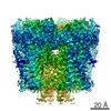 8354MC 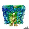 8355C 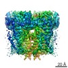 8356C M: map data used to model this data C: citing same article ( |
|---|---|
| Similar structure data |


-Links
 PDBj
PDBj

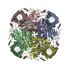
- Assembly
| Deposited unit | 
|
|---|---|
| 1 |
|
-Components
| #1: Protein | Mass: 59429.145 Da / Num. of mol.: 4 Source method: isolated from a genetically manipulated source Source: (gene. exp.) Homo sapiens (human) / Gene: PKD2, TRPP2 / Cell line (production host): HEK293S GnTI-/- / Production host: Homo sapiens (human) / References: UniProt: Q13563#2: Sugar | ChemComp-NAG /   Type: D-saccharide, beta linking / Mass: 221.208 Da / Num. of mol.: 12 Type: D-saccharide, beta linking / Mass: 221.208 Da / Num. of mol.: 12Source method: isolated from a genetically manipulated source Formula: C8H15NO6 Has protein modification | Y | |
|---|
-Experimental details
-Experiment
| Experiment | Method: ELECTRON MICROSCOPY |
|---|---|
| EM experiment | Aggregation state: PARTICLE / 3D reconstruction method: single particle reconstruction |
- Sample preparation
| Component | Name: hPKD:198-703 / Type: COMPLEX / Entity ID: #1 / Source: RECOMBINANT | |||||||||||||||
|---|---|---|---|---|---|---|---|---|---|---|---|---|---|---|---|---|
| Source (natural) | Organism: Homo sapiens (human) | |||||||||||||||
| Source (recombinant) | Organism: Homo sapiens (human) / Cell: HEK293S GnTI-/- / Plasmid: pFastbac1 | |||||||||||||||
| Buffer solution | pH: 7.4 | |||||||||||||||
| Buffer component |
| |||||||||||||||
| Specimen | Conc.: 2 mg/ml / Embedding applied: NO / Shadowing applied: NO / Staining applied: NO / Vitrification applied: YES Details: Single particles embedded in lipid nanodiscs. This sample was monodisperse. | |||||||||||||||
| Specimen support | Grid material: COPPER / Grid mesh size: 400 divisions/in. / Grid type: Quantifoil | |||||||||||||||
| Vitrification | Instrument: FEI VITROBOT MARK II / Cryogen name: ETHANE / Humidity: 80 % / Chamber temperature: 277 K / Details: blot for 7 seconds, -1 mm offset before plunging |
- Electron microscopy imaging
| Experimental equipment |  Model: Tecnai Polara / Image courtesy: FEI Company |
|---|---|
| Microscopy | Model: FEI POLARA 300 |
| Electron gun | Electron source:  FIELD EMISSION GUN / Accelerating voltage: 300 kV / Illumination mode: FLOOD BEAM FIELD EMISSION GUN / Accelerating voltage: 300 kV / Illumination mode: FLOOD BEAM |
| Electron lens | Mode: BRIGHT FIELD / Nominal magnification: 31000 X / Calibrated magnification: 41132 X / Nominal defocus max: 2400 nm / Nominal defocus min: 600 nm / Cs: 2 mm |
| Specimen holder | Cryogen: NITROGEN |
| Image recording | Average exposure time: 0.2 sec. / Electron dose: 1.35 e/Å2 / Detector mode: SUPER-RESOLUTION / Film or detector model: GATAN K2 SUMMIT (4k x 4k) / Num. of grids imaged: 1 / Num. of real images: 1500 |
| Image scans | Movie frames/image: 40 |
- Processing
| Software | Name: PHENIX / Version: 1.10.1_2155: / Classification: refinement | ||||||||||||||||||||||||||||||||||||
|---|---|---|---|---|---|---|---|---|---|---|---|---|---|---|---|---|---|---|---|---|---|---|---|---|---|---|---|---|---|---|---|---|---|---|---|---|---|
| EM software |
| ||||||||||||||||||||||||||||||||||||
| Image processing | Details: superresolution mode | ||||||||||||||||||||||||||||||||||||
| CTF correction | Type: PHASE FLIPPING AND AMPLITUDE CORRECTION | ||||||||||||||||||||||||||||||||||||
| Particle selection | Num. of particles selected: 368032 / Details: semi-automated particle picking | ||||||||||||||||||||||||||||||||||||
| Symmetry | Point symmetry: C4 (4 fold cyclic) | ||||||||||||||||||||||||||||||||||||
| 3D reconstruction | Resolution: 3 Å / Resolution method: FSC 0.143 CUT-OFF / Num. of particles: 93805 / Algorithm: FOURIER SPACE / Num. of class averages: 1 / Symmetry type: POINT | ||||||||||||||||||||||||||||||||||||
| Refine LS restraints |
|
