 Movie
Movie Controller


+ Open data

- Basic information
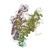
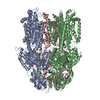
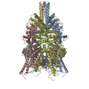
| Entry | Database: PDB / ID: 5yx9 | ||||||
|---|---|---|---|---|---|---|---|
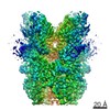
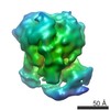
| Title | Cryo-EM structure of human TRPC6 at 3.8A resolution | ||||||
 Components Components | Short transient receptor potential channel 6 | ||||||
 Keywords Keywords | MEMBRANE PROTEIN / TRPC6 channel | ||||||
| Function / homology |  Function and homology information Function and homology informationpositive regulation of ion transmembrane transporter activity / slit diaphragm / negative regulation of dendrite morphogenesis / Role of second messengers in netrin-1 signaling / store-operated calcium channel activity / Effects of PIP2 hydrolysis / Elevation of cytosolic Ca2+ levels / cation channel complex / positive regulation of calcium ion transport / inositol 1,4,5 trisphosphate binding ...positive regulation of ion transmembrane transporter activity / slit diaphragm / negative regulation of dendrite morphogenesis / Role of second messengers in netrin-1 signaling / store-operated calcium channel activity / Effects of PIP2 hydrolysis / Elevation of cytosolic Ca2+ levels / cation channel complex / positive regulation of calcium ion transport / inositol 1,4,5 trisphosphate binding / actinin binding / clathrin binding / single fertilization / TRP channels / monoatomic cation transport / regulation of cytosolic calcium ion concentration / monoatomic cation channel activity / positive regulation of neuron differentiation / cellular response to hydrogen peroxide / neuron differentiation / calcium channel activity / calcium ion transmembrane transport / positive regulation of cytosolic calcium ion concentration / ATPase binding / actin binding / cellular response to hypoxia / protein homodimerization activity / membrane / plasma membrane / cytoplasm Similarity search - Function | ||||||
| Biological species |  Homo sapiens (human) Homo sapiens (human) | ||||||
| Method | ELECTRON MICROSCOPY / single particle reconstruction / cryo EM / Resolution: 3.8 Å | ||||||
 Authors Authors | Chen, L. / Tang, Q. / Guo, W. | ||||||
 Citation Citation | Journal: Cell Res / Year: 2018 Title: Structure of the receptor-activated human TRPC6 and TRPC3 ion channels. Authors: Qinglin Tang / Wenjun Guo / Li Zheng / Jing-Xiang Wu / Meng Liu / Xindi Zhou / Xiaolin Zhang / Lei Chen /  Abstract: TRPC6 and TRPC3 are receptor-activated nonselective cation channels that belong to the family of canonical transient receptor potential (TRPC) channels. They are activated by diacylglycerol, a lipid ...TRPC6 and TRPC3 are receptor-activated nonselective cation channels that belong to the family of canonical transient receptor potential (TRPC) channels. They are activated by diacylglycerol, a lipid second messenger. TRPC6 and TRPC3 are involved in many physiological processes and implicated in human genetic diseases. Here we present the structure of human TRPC6 homotetramer in complex with a newly identified high-affinity inhibitor BTDM solved by single-particle cryo-electron microscopy to 3.8 Å resolution. We also present the structure of human TRPC3 at 4.4 Å resolution. These structures show two-layer architectures in which the bell-shaped cytosolic layer holds the transmembrane layer. Extensive inter-subunit interactions of cytosolic domains, including the N-terminal ankyrin repeats and the C-terminal coiled-coil, contribute to the tetramer assembly. The high-affinity inhibitor BTDM wedges between the S5-S6 pore domain and voltage sensor-like domain to inhibit channel opening. Our structures uncover the molecular architecture of TRPC channels and provide a structural basis for understanding the mechanism of these channels. | ||||||
| History |
|
- Structure visualization
| Movie |
Movie viewer |
|---|---|
| Structure viewer | Molecule: MolmilJmol/JSmol |
- Downloads & links
-Download
| PDBx/mmCIF format | 5yx9.cif.gz | 538.9 KB | Display | PDBx/mmCIF format |
|---|---|---|---|---|
| PDB format | pdb5yx9.ent.gz | 434.2 KB | Display | PDB format |
| PDBx/mmJSON format | 5yx9.json.gz | Tree view | PDBx/mmJSON format | |
| Others |  Other downloads Other downloads |
-Validation report
| Arichive directory | https://data.pdbj.org/pub/pdb/validation_reports/yx/5yx9ftp://data.pdbj.org/pub/pdb/validation_reports/yx/5yx9 | HTTPS FTP |
|---|
-Related structure data
| Related structure data |  6856MC 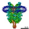 6911C 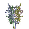 5zbgC M: map data used to model this data C: citing same article ( |
|---|---|
| Similar structure data |


-Links
 PDBj
PDBj
- Assembly
| Deposited unit | 
|
|---|---|
| 1 |
|
-Components
| #1: Protein | Mass: 106453.242 Da / Num. of mol.: 4 Source method: isolated from a genetically manipulated source Source: (gene. exp.) Homo sapiens (human) / Production host: Homo sapiens (human) / References: UniProt: Q9Y210 |
|---|
-Experimental details
-Experiment
| Experiment | Method: ELECTRON MICROSCOPY |
|---|---|
| EM experiment | Aggregation state: PARTICLE / 3D reconstruction method: single particle reconstruction |
- Sample preparation
| Component | Name: hTRPC6 homo-tetramer in complex with inhibitor / Type: ORGANELLE OR CELLULAR COMPONENT / Entity ID: all / Source: RECOMBINANT |
|---|---|
| Source (natural) | Organism: Homo sapiens (human) |
| Source (recombinant) | Organism: Homo sapiens (human) |
| Buffer solution | pH: 7.5 |
| Specimen | Embedding applied: NO / Shadowing applied: NO / Staining applied: NO / Vitrification applied: YES |
| Vitrification | Cryogen name: ETHANE |
- Electron microscopy imaging
| Experimental equipment |  Model: Titan Krios / Image courtesy: FEI Company |
|---|---|
| Microscopy | Model: FEI TITAN KRIOS |
| Electron gun | Electron source:  FIELD EMISSION GUN / Accelerating voltage: 300 kV / Illumination mode: FLOOD BEAM FIELD EMISSION GUN / Accelerating voltage: 300 kV / Illumination mode: FLOOD BEAM |
| Electron lens | Mode: BRIGHT FIELD |
| Image recording | Electron dose: 50 e/Å2 / Film or detector model: GATAN K2 SUMMIT (4k x 4k) |
- Processing
| EM software |
| |||||||||
|---|---|---|---|---|---|---|---|---|---|---|
| CTF correction | Type: NONE | |||||||||
| Symmetry | Point symmetry: C4 (4 fold cyclic) | |||||||||
| 3D reconstruction | Resolution: 3.8 Å / Resolution method: FSC 0.143 CUT-OFF / Num. of particles: 53528 / Symmetry type: POINT |
