 Movie
Movie Controller

[English] 日本語
 Yorodumi
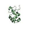
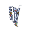
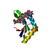
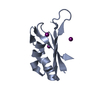
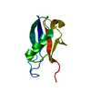
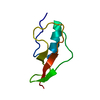
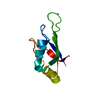
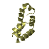
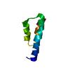
- PDB-6a6c: Crystal structure of carbohydrate-binding module family 56 beta-1... -
+ Open data

- Basic information
| Entry | Database: PDB / ID: 6a6c | ||||||
|---|---|---|---|---|---|---|---|
| Title | Crystal structure of carbohydrate-binding module family 56 beta-1,3-glucan binding domain | ||||||
 Components Components | Beta-1,3-glucanase | ||||||
 Keywords Keywords | SUGAR BINDING PROTEIN / carbohydrate-binding module family 56 / beta-1 / 3-glucan / sugar binding | ||||||
| Function / homology |  Function and homology information Function and homology informationglucan endo-1,3-beta-D-glucosidase / glucan endo-1,3-beta-D-glucosidase activity / carbohydrate binding Similarity search - Function | ||||||
| Biological species |  Paenibacillus barengoltzii (bacteria) Paenibacillus barengoltzii (bacteria) | ||||||
| Method |  X-RAY DIFFRACTION / SYNCHROTRON / MOLECULAR REPLACEMENT / Resolution: 2.05 Å X-RAY DIFFRACTION / SYNCHROTRON / MOLECULAR REPLACEMENT / Resolution: 2.05 Å | ||||||
 Authors Authors | Qin, Z. / Lin, S. | ||||||
| Funding support |  China, 1items China, 1items
| ||||||
 Citation Citation | Journal: To Be Published Title: Crystal structure of carbohydrate-binding module family 56 beta-1,3-glucan binding domain Authors: Qin, Z. / Lin, S. | ||||||
| History |
|
- Structure visualization
| Structure viewer | Molecule: MolmilJmol/JSmol |
|---|
- Downloads & links
-Download
| PDBx/mmCIF format | 6a6c.cif.gz | 28.9 KB | Display | PDBx/mmCIF format |
|---|---|---|---|---|
| PDB format | pdb6a6c.ent.gz | 17.9 KB | Display | PDB format |
| PDBx/mmJSON format | 6a6c.json.gz | Tree view | PDBx/mmJSON format | |
| Others |  Other downloads Other downloads |
-Validation report
| Arichive directory | https://data.pdbj.org/pub/pdb/validation_reports/a6/6a6cftp://data.pdbj.org/pub/pdb/validation_reports/a6/6a6c | HTTPS FTP |
|---|
-Related structure data
| Related structure data | 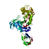 5h9xS S: Starting model for refinement |
|---|---|
| Similar structure data |

-Links
 PDBj
PDBj
- Assembly
| Deposited unit | 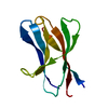
| ||||||||
|---|---|---|---|---|---|---|---|---|---|
| 1 |
| ||||||||
| Unit cell |
|
-Components
| #1: Protein | Mass: 9561.397 Da / Num. of mol.: 1 Source method: isolated from a genetically manipulated source Details: SF file contains Friedel pairs. / Source: (gene. exp.) Paenibacillus barengoltzii (bacteria)Production host: References: UniProt: A0A1S4NYE1, glucan endo-1,3-beta-D-glucosidase |
|---|---|
| #2: Water | ChemComp-HOH /  Mass: 18.015 Da / Num. of mol.: 2 / Source method: isolated from a natural source / Formula: H2O Mass: 18.015 Da / Num. of mol.: 2 / Source method: isolated from a natural source / Formula: H2O |
-Experimental details
-Experiment
| Experiment | Method: X-RAY DIFFRACTION / Number of used crystals: 1 |
|---|
- Sample preparation
| Crystal | Density Matthews: 2.23 Å3/Da / Density % sol: 44.8 % |
|---|---|
| Crystal grow | Temperature: 293.15 K / Method: vapor diffusion, sitting drop / pH: 7 Details: 14 mM sodium cholate,1.4 M di-ammonium tartrate, 20 days |
-Data collection
| Diffraction | Mean temperature: 100 K |
|---|---|
| Diffraction source | Source: SYNCHROTRON / Site: SSRF / Beamline: BL17U1 / Wavelength: 1.0093 Å |
| Detector | Type: MARMOSAIC 225 mm CCD / Detector: CCD / Date: Oct 16, 2017 |
| Radiation | Protocol: SINGLE WAVELENGTH / Monochromatic (M) / Laue (L): M / Scattering type: x-ray |
| Radiation wavelength | Wavelength: 1.0093 Å / Relative weight: 1 |
| Reflection | Resolution: 2.05→33.712 Å / Num. obs: 8208 / % possible obs: 87.68 % / Redundancy: 18.5 % / Rmerge(I) obs: 0.105 / Net I/σ(I): 25.86 |
| Reflection shell | Resolution: 2.05→2.123 Å / Rmerge(I) obs: 0.412 / Num. unique obs: 537 |
- Processing
| Software |
| ||||||||||||||||||||||||||||||||||||||||||
|---|---|---|---|---|---|---|---|---|---|---|---|---|---|---|---|---|---|---|---|---|---|---|---|---|---|---|---|---|---|---|---|---|---|---|---|---|---|---|---|---|---|---|---|
| Refinement | Method to determine structure: MOLECULAR REPLACEMENT Starting model: 5H9X Resolution: 2.05→29.472 Å / SU ML: 0.37 / Cross valid method: FREE R-VALUE / σ(F): 1.35 / Phase error: 26.97
| ||||||||||||||||||||||||||||||||||||||||||
| Solvent computation | Shrinkage radii: 0.9 Å / VDW probe radii: 1.11 Å | ||||||||||||||||||||||||||||||||||||||||||
| Refinement step | Cycle: LAST / Resolution: 2.05→29.472 Å
| ||||||||||||||||||||||||||||||||||||||||||
| Refine LS restraints |
| ||||||||||||||||||||||||||||||||||||||||||
| LS refinement shell |
|
