 Movie
Movie Controller

[English] 日本語
 Yorodumi
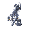
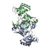
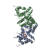
- PDB-5h9x: Crystal structure of GH family 64 laminaripentaose-producing beta... -
+ Open data

- Basic information
| Entry | Database: PDB / ID: 5h9x | ||||||
|---|---|---|---|---|---|---|---|
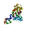
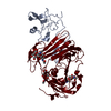
| Title | Crystal structure of GH family 64 laminaripentaose-producing beta-1,3-glucanase from Paenibacillus barengoltzii | ||||||
 Components Components | beta-1,3-glucanase | ||||||
 Keywords Keywords | HYDROLASE / beta-1 / 3-glucan recognition / glycoside hydrolase family 64 / 3-glucanase / GH64-TLP-SF superfamily | ||||||
| Function / homology |  Function and homology information Function and homology informationglucan endo-1,3-beta-D-glucosidase / glucan endo-1,3-beta-D-glucosidase activity / carbohydrate binding Similarity search - Function | ||||||
| Biological species |  Paenibacillus barengoltzii (bacteria) Paenibacillus barengoltzii (bacteria) | ||||||
| Method |  X-RAY DIFFRACTION / SYNCHROTRON / SAD / Resolution: 1.91 Å X-RAY DIFFRACTION / SYNCHROTRON / SAD / Resolution: 1.91 Å | ||||||
 Authors Authors | Zhen, Q. / Yan, Q. / Yang, S. / Jiang, Z. / You, X. | ||||||
 Citation Citation | Journal: Chem. Commun. (Camb.) / Year: 2017 Title: The recognition mechanism of triple-helical beta-1,3-glucan by a beta-1,3-glucanase Authors: Qin, Z. / Yang, D. / You, X. / Liu, Y. / Hu, S. / Yan, Q. / Yang, S. / Jiang, Z. | ||||||
| History |
|
- Structure visualization
| Structure viewer | Molecule: MolmilJmol/JSmol |
|---|
- Downloads & links
-Download
| PDBx/mmCIF format | 5h9x.cif.gz | 103.4 KB | Display | PDBx/mmCIF format |
|---|---|---|---|---|
| PDB format | pdb5h9x.ent.gz | 76.8 KB | Display | PDB format |
| PDBx/mmJSON format | 5h9x.json.gz | Tree view | PDBx/mmJSON format | |
| Others |  Other downloads Other downloads |
-Validation report
| Arichive directory | https://data.pdbj.org/pub/pdb/validation_reports/h9/5h9xftp://data.pdbj.org/pub/pdb/validation_reports/h9/5h9x | HTTPS FTP |
|---|
-Related structure data


-Links
 PDBj
PDBj
- Assembly
| Deposited unit | 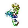
| ||||||||
|---|---|---|---|---|---|---|---|---|---|
| 1 |
| ||||||||
| Unit cell |
|
-Components
| #1: Protein | Mass: 49075.855 Da / Num. of mol.: 1 Source method: isolated from a genetically manipulated source Source: (gene. exp.) Paenibacillus barengoltzii / Strain: CAU904 Production host: References: UniProt: A0A1S4NYE1*PLUS, glucan endo-1,3-beta-D-glucosidase |
|---|---|
| #2: Water | ChemComp-HOH /  Mass: 18.015 Da / Num. of mol.: 347 / Source method: isolated from a natural source / Formula: H2O Mass: 18.015 Da / Num. of mol.: 347 / Source method: isolated from a natural source / Formula: H2O |
| Sequence details | AUTHORS STATE THAT THE GENEBANK ACCESSION NUMBER IS KU363233 FOR THIS SEQUENCE. |
-Experimental details
-Experiment
| Experiment | Method: X-RAY DIFFRACTION |
|---|
- Sample preparation
| Crystal | Density Matthews: 2.81 Å3/Da / Density % sol: 56.25 % |
|---|---|
| Crystal grow | Temperature: 293 K / Method: vapor diffusion, sitting drop / pH: 7 Details: Optimized crystals suitable for diffraction were grown in drops containing 2 microliter of protein solution and 0.5 microliter of reservoir solution (1.2M di-Ammonium Tartrate pH 7.0) at 293 ...Details: Optimized crystals suitable for diffraction were grown in drops containing 2 microliter of protein solution and 0.5 microliter of reservoir solution (1.2M di-Ammonium Tartrate pH 7.0) at 293 K. The crystals were observed 10 days later. PH range: 7 |
-Data collection
| Diffraction | Mean temperature: 100 K |
|---|---|
| Diffraction source | Source: SYNCHROTRON / Site: Photon Factory  / Beamline: BL-1A / Wavelength: 1.1 Å / Beamline: BL-1A / Wavelength: 1.1 Å |
| Detector | Type: ADSC QUANTUM 315 / Detector: CCD / Date: Dec 15, 2014 |
| Radiation | Protocol: SINGLE WAVELENGTH / Monochromatic (M) / Laue (L): M / Scattering type: x-ray |
| Radiation wavelength | Wavelength: 1.1 Å / Relative weight: 1 |
| Reflection | Resolution: 1.91→32.32 Å / Num. obs: 36956 / % possible obs: 91.16 % / Redundancy: 6.3 % / Rmerge(I) obs: 0.095 / Rsym value: 0.052 / Net I/σ(I): 13.72 |
| Reflection shell | Resolution: 1.91→1.97 Å / % possible all: 89.91 |
- Processing
| Software |
| |||||||||||||||||||||||||||||||||||||||||||||||||||||||||||||||||||||||||||||||||||||||||||||||||||||||||
|---|---|---|---|---|---|---|---|---|---|---|---|---|---|---|---|---|---|---|---|---|---|---|---|---|---|---|---|---|---|---|---|---|---|---|---|---|---|---|---|---|---|---|---|---|---|---|---|---|---|---|---|---|---|---|---|---|---|---|---|---|---|---|---|---|---|---|---|---|---|---|---|---|---|---|---|---|---|---|---|---|---|---|---|---|---|---|---|---|---|---|---|---|---|---|---|---|---|---|---|---|---|---|---|---|---|---|
| Refinement | Method to determine structure: SAD / Resolution: 1.91→32.317 Å / SU ML: 0.22 / Cross valid method: FREE R-VALUE / σ(F): 1.34 / Phase error: 25.44 / Stereochemistry target values: ML
| |||||||||||||||||||||||||||||||||||||||||||||||||||||||||||||||||||||||||||||||||||||||||||||||||||||||||
| Solvent computation | Shrinkage radii: 0.9 Å / VDW probe radii: 1.11 Å / Solvent model: FLAT BULK SOLVENT MODEL | |||||||||||||||||||||||||||||||||||||||||||||||||||||||||||||||||||||||||||||||||||||||||||||||||||||||||
| Refinement step | Cycle: LAST / Resolution: 1.91→32.317 Å
| |||||||||||||||||||||||||||||||||||||||||||||||||||||||||||||||||||||||||||||||||||||||||||||||||||||||||
| Refine LS restraints |
| |||||||||||||||||||||||||||||||||||||||||||||||||||||||||||||||||||||||||||||||||||||||||||||||||||||||||
| LS refinement shell |
|
