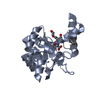
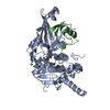
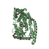
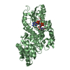
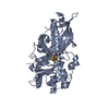
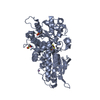
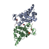
Entry Database : PDB / ID : 6a6aTitle VanYB in complex with D-Alanine D-alanyl-D-alanine carboxypeptidase Keywords / / / Function / homology Function Domain/homology Component
/ / / / / / / / / / / / / / / / Biological species Enterococcus faecalis (bacteria)Method / / / Resolution : 2.26 Å Authors Kim, H.S. / Hahn, H. Funding support Organization Grant number Country Ministry of Science, ICT and Future Planning 2013R1A2A1A05067303 Ministry of Science, ICT and Future Planning NRF-2011-0030001 Ministry of Science, ICT and Future Planning NRF-2013M3A6A4043695 Ministry of Science, ICT and Future Planning NRF-2017R1C1B2012225
Journal : Int. J. Biol. Macromol. / Year : 2018Title : Structural basis for the substrate recognition of peptidoglycan pentapeptides by Enterococcus faecalis VanYB.Authors : Kim, H.S. / Hahn, H. / Kim, J. / Jang, D.M. / Lee, J.Y. / Back, J.M. / Im, H.N. / Kim, H. / Han, B.W. / Suh, S.W. History Deposition Jun 27, 2018 Deposition site / Processing site Revision 1.0 Sep 5, 2018 Provider / Type Revision 1.1 Nov 22, 2023 Group Data collection / Database references ... Data collection / Database references / Derived calculations / Refinement description Category chem_comp_atom / chem_comp_bond ... chem_comp_atom / chem_comp_bond / database_2 / pdbx_initial_refinement_model / struct_conn Item _database_2.pdbx_DOI / _database_2.pdbx_database_accession ... _database_2.pdbx_DOI / _database_2.pdbx_database_accession / _struct_conn.pdbx_dist_value / _struct_conn.ptnr1_auth_asym_id / _struct_conn.ptnr1_auth_comp_id / _struct_conn.ptnr1_auth_seq_id / _struct_conn.ptnr1_label_asym_id / _struct_conn.ptnr1_label_atom_id / _struct_conn.ptnr1_label_comp_id / _struct_conn.ptnr1_label_seq_id / _struct_conn.ptnr2_auth_asym_id / _struct_conn.ptnr2_label_asym_id
Show all Show less
 Movie
Movie Controller


 Open data
 Basic information
 Components
 Keywords
 Function and homology information
 Enterococcus faecalis (bacteria)
 X-RAY DIFFRACTION /
  Authors
 Korea, Republic Of, 4items
  Citation
 Structure visualization
 Downloads & links
 Other downloads
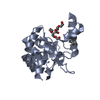


 PDBj
PDBj
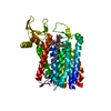
 Assembly
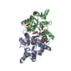
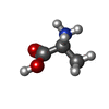


 Type: D-peptide linking / Mass: 89.093 Da / Num. of mol.: 2 / Source method: isolated from a natural source / Formula: C3H7NO2 / Feature type: SUBJECT OF INVESTIGATION
 Mass: 92.094 Da / Num. of mol.: 4 / Source method: obtained synthetically / Formula: C3H8O3
 Mass: 59.044 Da / Num. of mol.: 2 / Source method: obtained synthetically / Formula: C2H3O2
 Mass: 65.409 Da / Num. of mol.: 2 / Source method: obtained synthetically / Formula: Zn
 Mass: 150.173 Da / Num. of mol.: 4 / Source method: obtained synthetically / Formula: C6H14O4
 Sample preparation
 Processing