 Movie
Movie Controller

[English] 日本語
 Yorodumi
- PDB-6fwz: Crystal structure of human UDP-N-acetylglucosamine-dolichyl-phosp... -
+ Open data

- Basic information
| Entry | Database: PDB / ID: 6fwz | ||||||
|---|---|---|---|---|---|---|---|
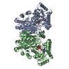
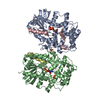
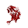
| Title | Crystal structure of human UDP-N-acetylglucosamine-dolichyl-phosphate N-acetylglucosaminephosphotransferase (DPAGT1) (V264G mutant) in complex with UDP-GlcNAc | ||||||
 Components Components | UDP-N-acetylglucosamine--dolichyl-phosphate N-acetylglucosaminephosphotransferase | ||||||
 Keywords Keywords | TRANSFERASE / Protein glycosylation / integral membrane protein / PNPT / congenital myasthenic syndrome / antibiotic / Structural Genomics / Structural Genomics Consortium / SGC | ||||||
| Function / homology |  Function and homology information Function and homology informationDefective DPAGT1 causes CDG-1j, CMSTA2 / UDP-N-acetylglucosamine-dolichyl-phosphate N-acetylglucosaminephosphotransferase / UDP-N-acetylglucosamine-dolichyl-phosphate N-acetylglucosaminephosphotransferase activity / Biosynthesis of the N-glycan precursor (dolichol lipid-linked oligosaccharide, LLO) and transfer to a nascent protein / UDP-N-acetylglucosamine-lysosomal-enzyme N-acetylglucosaminephosphotransferase activity / dolichol-linked oligosaccharide biosynthetic process / protein N-linked glycosylation / glycosyltransferase activity / intracellular membrane-bounded organelle / endoplasmic reticulum membrane ...Defective DPAGT1 causes CDG-1j, CMSTA2 / UDP-N-acetylglucosamine-dolichyl-phosphate N-acetylglucosaminephosphotransferase / UDP-N-acetylglucosamine-dolichyl-phosphate N-acetylglucosaminephosphotransferase activity / Biosynthesis of the N-glycan precursor (dolichol lipid-linked oligosaccharide, LLO) and transfer to a nascent protein / UDP-N-acetylglucosamine-lysosomal-enzyme N-acetylglucosaminephosphotransferase activity / dolichol-linked oligosaccharide biosynthetic process / protein N-linked glycosylation / glycosyltransferase activity / intracellular membrane-bounded organelle / endoplasmic reticulum membrane / Golgi apparatus / membrane / metal ion binding / identical protein binding Similarity search - Function | ||||||
| Biological species |  Homo sapiens (human) Homo sapiens (human) | ||||||
| Method |  X-RAY DIFFRACTION / SYNCHROTRON / MOLECULAR REPLACEMENT / Resolution: 3.1 Å X-RAY DIFFRACTION / SYNCHROTRON / MOLECULAR REPLACEMENT / Resolution: 3.1 Å | ||||||
 Authors Authors | Pike, A.C.W. / Dong, Y.Y. / Chu, A. / Tessitore, A. / Goubin, S. / Dong, L. / Mukhopadhyay, S. / Mahajan, P. / Chalk, R. / Berridge, G. ...Pike, A.C.W. / Dong, Y.Y. / Chu, A. / Tessitore, A. / Goubin, S. / Dong, L. / Mukhopadhyay, S. / Mahajan, P. / Chalk, R. / Berridge, G. / Wang, D. / Kupinska, K. / Belaya, K. / Beeson, D. / Burgess-Brown, N. / Edwards, A.M. / Arrowsmith, C.H. / Bountra, C. / Carpenter, E.P. / Structural Genomics Consortium (SGC) | ||||||
| Funding support |  United Kingdom, 1items United Kingdom, 1items
| ||||||
 Citation Citation | Journal: Cell / Year: 2018 Title: Structures of DPAGT1 Explain Glycosylation Disease Mechanisms and Advance TB Antibiotic Design. Authors: Dong, Y.Y. / Wang, H. / Pike, A.C.W. / Cochrane, S.A. / Hamedzadeh, S. / Wyszynski, F.J. / Bushell, S.R. / Royer, S.F. / Widdick, D.A. / Sajid, A. / Boshoff, H.I. / Park, Y. / Lucas, R. / ...Authors: Dong, Y.Y. / Wang, H. / Pike, A.C.W. / Cochrane, S.A. / Hamedzadeh, S. / Wyszynski, F.J. / Bushell, S.R. / Royer, S.F. / Widdick, D.A. / Sajid, A. / Boshoff, H.I. / Park, Y. / Lucas, R. / Liu, W.M. / Lee, S.S. / Machida, T. / Minall, L. / Mehmood, S. / Belaya, K. / Liu, W.W. / Chu, A. / Shrestha, L. / Mukhopadhyay, S.M.M. / Strain-Damerell, C. / Chalk, R. / Burgess-Brown, N.A. / Bibb, M.J. / Barry Iii, C.E. / Robinson, C.V. / Beeson, D. / Davis, B.G. / Carpenter, E.P. | ||||||
| History |
|
- Structure visualization
| Structure viewer | Molecule: MolmilJmol/JSmol |
|---|
- Downloads & links
-Download
| PDBx/mmCIF format | 6fwz.cif.gz | 170.4 KB | Display | PDBx/mmCIF format |
|---|---|---|---|---|
| PDB format | pdb6fwz.ent.gz | 134.7 KB | Display | PDB format |
| PDBx/mmJSON format | 6fwz.json.gz | Tree view | PDBx/mmJSON format | |
| Others |  Other downloads Other downloads |
-Validation report
| Arichive directory | https://data.pdbj.org/pub/pdb/validation_reports/fw/6fwzftp://data.pdbj.org/pub/pdb/validation_reports/fw/6fwz | HTTPS FTP |
|---|
-Related structure data
| Related structure data |  5levSC  5o5eC  6fm9C S: Starting model for refinement C: citing same article ( |
|---|---|
| Similar structure data |


-Links
 PDBj
PDBj

- Assembly
| Deposited unit | 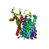
| ||||||||
|---|---|---|---|---|---|---|---|---|---|
| 1 | 
| ||||||||
| Unit cell |
|
-Components
-Protein , 1 types, 1 molecules A
| #1: Protein | Mass: 46173.719 Da / Num. of mol.: 1 / Mutation: V264G Source method: isolated from a genetically manipulated source Source: (gene. exp.) Homo sapiens (human) / Gene: DPAGT1, DPAGT2 / Plasmid: PFB-LIC-BSE / Production host:   Spodoptera frugiperda (fall armyworm) Spodoptera frugiperda (fall armyworm)References: UniProt: Q9H3H5, UDP-N-acetylglucosamine-dolichyl-phosphate N-acetylglucosaminephosphotransferase |
|---|
-Non-polymers , 5 types, 23 molecules 


| #2: Chemical | ChemComp-MG /  Mass: 24.305 Da / Num. of mol.: 1 / Source method: obtained synthetically / Formula: Mg Mass: 24.305 Da / Num. of mol.: 1 / Source method: obtained synthetically / Formula: Mg |
|---|---|
| #3: Chemical | ChemComp-UD1 /  Mass: 607.354 Da / Num. of mol.: 1 / Source method: obtained synthetically / Formula: C17H27N3O17P2 Mass: 607.354 Da / Num. of mol.: 1 / Source method: obtained synthetically / Formula: C17H27N3O17P2 |
| #4: Chemical | ChemComp-UNL / Num. of mol.: 1 / Source method: obtained synthetically |
| #5: Chemical | ChemComp-P6L / ( Mass: 746.991 Da / Num. of mol.: 1 / Source method: obtained synthetically / Formula: C40H75O10P / Comment: phospholipid*YM Mass: 746.991 Da / Num. of mol.: 1 / Source method: obtained synthetically / Formula: C40H75O10P / Comment: phospholipid*YM |
| #6: Water | ChemComp-HOH / Mass: 18.015 Da / Num. of mol.: 19 / Source method: isolated from a natural source / Formula: H2O |
-Experimental details
-Experiment
| Experiment | Method: X-RAY DIFFRACTION / Number of used crystals: 1 |
|---|
- Sample preparation
| Crystal | Density Matthews: 3.91 Å3/Da / Density % sol: 68.59 % |
|---|---|
| Crystal grow | Temperature: 293 K / Method: vapor diffusion, sitting drop / pH: 9 Details: 0.1M bicine pH 9.0 -- 0.05M sodium chloride -- 37.5% PEG300 |
-Data collection
| Diffraction | Mean temperature: 100 K |
|---|---|
| Diffraction source | Source: SYNCHROTRON / Site: Diamond / Beamline: I24 / Wavelength: 0.9686 Å |
| Detector | Type: DECTRIS PILATUS3 6M / Detector: PIXEL / Date: Jan 16, 2016 |
| Radiation | Protocol: SINGLE WAVELENGTH / Monochromatic (M) / Laue (L): M / Scattering type: x-ray |
| Radiation wavelength | Wavelength: 0.9686 Å / Relative weight: 1 |
| Reflection | Resolution: 3.1→59.55 Å / Num. obs: 14197 / % possible obs: 100 % / Redundancy: 9.5 % / Biso Wilson estimate: 85.75 Å2 / CC1/2: 0.999 / Rmerge(I) obs: 0.095 / Rpim(I) all: 0.033 / Rrim(I) all: 0.101 / Net I/σ(I): 14.4 |
| Reflection shell | Resolution: 3.1→3.18 Å / Redundancy: 9.9 % / Rmerge(I) obs: 2.376 / Mean I/σ(I) obs: 1.1 / Num. unique obs: 1002 / CC1/2: 0.387 / Rpim(I) all: 0.784 / Rrim(I) all: 2.505 / % possible all: 100 |
- Processing
| Software |
| ||||||||||||||||||||||||||||||||||||||||||||||||||||||||||||||||||||||||||||||||||||||||||||||||||||||||||||||||||||||||||||||||||||||||||||||||||||||||||||||||||||||||||||||||||||||
|---|---|---|---|---|---|---|---|---|---|---|---|---|---|---|---|---|---|---|---|---|---|---|---|---|---|---|---|---|---|---|---|---|---|---|---|---|---|---|---|---|---|---|---|---|---|---|---|---|---|---|---|---|---|---|---|---|---|---|---|---|---|---|---|---|---|---|---|---|---|---|---|---|---|---|---|---|---|---|---|---|---|---|---|---|---|---|---|---|---|---|---|---|---|---|---|---|---|---|---|---|---|---|---|---|---|---|---|---|---|---|---|---|---|---|---|---|---|---|---|---|---|---|---|---|---|---|---|---|---|---|---|---|---|---|---|---|---|---|---|---|---|---|---|---|---|---|---|---|---|---|---|---|---|---|---|---|---|---|---|---|---|---|---|---|---|---|---|---|---|---|---|---|---|---|---|---|---|---|---|---|---|---|---|
| Refinement | Method to determine structure: MOLECULAR REPLACEMENT Starting model: 5LEV Resolution: 3.1→30 Å / Cor.coef. Fo:Fc: 0.912 / Cor.coef. Fo:Fc free: 0.937 / SU B: 38.277 / SU ML: 0.314 / SU R Cruickshank DPI: 1.027 / Cross valid method: THROUGHOUT / ESU R: 1.027 / ESU R Free: 0.353 / SU Rfree Cruickshank DPI: 0.3529 Details: HYDROGENS HAVE BEEN ADDED IN THE RIDING POSITIONS. REFINED WITH AUTOBUSTER AND REFMAC IN FINAL CYCLE TO OPTIMISE UD1 GEOMETRY. PROSMART HBOND RESTRAINTS. SINGLE TLS GROUP FOR ENTIRE PROTEIN CHAIN
| ||||||||||||||||||||||||||||||||||||||||||||||||||||||||||||||||||||||||||||||||||||||||||||||||||||||||||||||||||||||||||||||||||||||||||||||||||||||||||||||||||||||||||||||||||||||
| Solvent computation | Ion probe radii: 0.8 Å / Shrinkage radii: 0.8 Å / VDW probe radii: 1.2 Å | ||||||||||||||||||||||||||||||||||||||||||||||||||||||||||||||||||||||||||||||||||||||||||||||||||||||||||||||||||||||||||||||||||||||||||||||||||||||||||||||||||||||||||||||||||||||
| Displacement parameters | Biso mean: 128.475 Å2
| ||||||||||||||||||||||||||||||||||||||||||||||||||||||||||||||||||||||||||||||||||||||||||||||||||||||||||||||||||||||||||||||||||||||||||||||||||||||||||||||||||||||||||||||||||||||
| Refinement step | Cycle: 1 / Resolution: 3.1→30 Å
| ||||||||||||||||||||||||||||||||||||||||||||||||||||||||||||||||||||||||||||||||||||||||||||||||||||||||||||||||||||||||||||||||||||||||||||||||||||||||||||||||||||||||||||||||||||||
| Refine LS restraints |
|