 Movie
Movie Controller

[English] 日本語
 Yorodumi
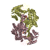
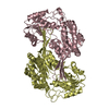
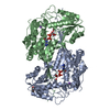
- PDB-3iwk: Crystal structure of aminoaldehyde dehydrogenase 1 from Pisum sat... -
+ Open data

- Basic information
| Entry | Database: PDB / ID: 3iwk | ||||||
|---|---|---|---|---|---|---|---|
| Title | Crystal structure of aminoaldehyde dehydrogenase 1 from Pisum sativum (PsAMADH1) | ||||||
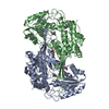
 Components Components | Aminoaldehyde dehydrogenase | ||||||
 Keywords Keywords | OXIDOREDUCTASE / Rossmann fold / dimer / aminoaldehyde dehydrogenase / betaine aldehyde dehydrogenase / NAD | ||||||
| Function / homology |  Function and homology information Function and homology informationgamma-guanidinobutyraldehyde dehydrogenase / gamma-guanidinobutyraldehyde dehydrogenase (NAD+) activity / 3-aminopropanal dehydrogenase (NAD+) activity / aminobutyraldehyde dehydrogenase / aminobutyraldehyde dehydrogenase (NAD+) activity / : / Oxidoreductases; Acting on the aldehyde or oxo group of donors; With NAD+ or NADP+ as acceptor / sodium ion binding / cellular detoxification of aldehyde / peroxisome ...gamma-guanidinobutyraldehyde dehydrogenase / gamma-guanidinobutyraldehyde dehydrogenase (NAD+) activity / 3-aminopropanal dehydrogenase (NAD+) activity / aminobutyraldehyde dehydrogenase / aminobutyraldehyde dehydrogenase (NAD+) activity / : / Oxidoreductases; Acting on the aldehyde or oxo group of donors; With NAD+ or NADP+ as acceptor / sodium ion binding / cellular detoxification of aldehyde / peroxisome / nucleotide binding / protein homodimerization activity Similarity search - Function | ||||||
| Biological species |  Pisum sativum (garden pea) Pisum sativum (garden pea) | ||||||
| Method |  X-RAY DIFFRACTION / SYNCHROTRON / MOLECULAR REPLACEMENT / molecular replacement / Resolution: 2.4 Å X-RAY DIFFRACTION / SYNCHROTRON / MOLECULAR REPLACEMENT / molecular replacement / Resolution: 2.4 Å | ||||||
 Authors Authors | Kopecny, D. / Morera, S. / Briozzo, P. | ||||||
 Citation Citation | Journal: J.Mol.Biol. / Year: 2010 Title: Structural and functional characterization of plant aminoaldehyde dehydrogenase from Pisum sativum with a broad specificity for natural and synthetic aminoaldehydes. Authors: Tylichova, M. / Kopecny, D. / Morera, S. / Briozzo, P. / Lenobel, R. / Snegaroff, J. / Sebela, M. | ||||||
| History |
|
- Structure visualization
| Structure viewer | Molecule: MolmilJmol/JSmol |
|---|
- Downloads & links
-Download
| PDBx/mmCIF format | 3iwk.cif.gz | 1.1 MB | Display | PDBx/mmCIF format |
|---|---|---|---|---|
| PDB format | pdb3iwk.ent.gz | 954.6 KB | Display | PDB format |
| PDBx/mmJSON format | 3iwk.json.gz | Tree view | PDBx/mmJSON format | |
| Others |  Other downloads Other downloads |
-Validation report
| Arichive directory | https://data.pdbj.org/pub/pdb/validation_reports/iw/3iwkftp://data.pdbj.org/pub/pdb/validation_reports/iw/3iwk | HTTPS FTP |
|---|
-Related structure data
| Related structure data | 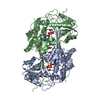 3iwjC 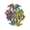 1cw3S C: citing same article ( S: Starting model for refinement |
|---|---|
| Similar structure data |


-Links
 PDBj
PDBj

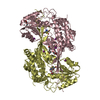
- Assembly
| Deposited unit | 
| ||||||||
|---|---|---|---|---|---|---|---|---|---|
| 1 | 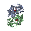
| ||||||||
| 2 | 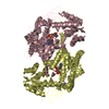
| ||||||||
| 3 | 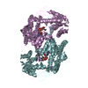
| ||||||||
| 4 | 
| ||||||||
| 5 | 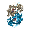
| ||||||||
| 6 | 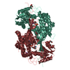
| ||||||||
| Unit cell |
|
-Components
-Protein , 1 types, 12 molecules ABCDEFGHIJKL
| #1: Protein | Mass: 54868.746 Da / Num. of mol.: 12 Source method: isolated from a genetically manipulated source Source: (gene. exp.) Pisum sativum (garden pea) / Gene: amadh1 / Plasmid: pET28 / Production host:  References: UniProt: Q8VWZ1, aminobutyraldehyde dehydrogenase |
|---|
-Non-polymers , 5 types, 2041 molecules 


| #2: Chemical | ChemComp-NA /  Mass: 22.990 Da / Num. of mol.: 12 / Source method: obtained synthetically / Formula: Na Mass: 22.990 Da / Num. of mol.: 12 / Source method: obtained synthetically / Formula: Na#3: Chemical | ChemComp-NAD /  Mass: 663.425 Da / Num. of mol.: 12 / Source method: obtained synthetically / Formula: C21H27N7O14P2 / Comment: NAD*YM Mass: 663.425 Da / Num. of mol.: 12 / Source method: obtained synthetically / Formula: C21H27N7O14P2 / Comment: NAD*YM#4: Chemical | ChemComp-GOL /  Mass: 92.094 Da / Num. of mol.: 19 / Source method: obtained synthetically / Formula: C3H8O3 Mass: 92.094 Da / Num. of mol.: 19 / Source method: obtained synthetically / Formula: C3H8O3#5: Chemical | ChemComp-MRD / ( |  Mass: 118.174 Da / Num. of mol.: 1 / Source method: obtained synthetically / Formula: C6H14O2 / Comment: precipitant*YM Mass: 118.174 Da / Num. of mol.: 1 / Source method: obtained synthetically / Formula: C6H14O2 / Comment: precipitant*YM#6: Water | ChemComp-HOH / | Mass: 18.015 Da / Num. of mol.: 1997 / Source method: isolated from a natural source / Formula: H2O |
|---|
-Experimental details
-Experiment
| Experiment | Method: X-RAY DIFFRACTION / Number of used crystals: 1 |
|---|
- Sample preparation
| Crystal | Density Matthews: 2.9 Å3/Da / Density % sol: 57.58 % |
|---|---|
| Crystal grow | Temperature: 293 K / Method: vapor diffusion, hanging drop / pH: 7.5 Details: 0.1M HEPES pH 7.5, 13% PEG 6000, 5% 2-methyl-2,4-pentanediol, 5mM NAD, VAPOR DIFFUSION, HANGING DROP, temperature 293K |
-Data collection
| Diffraction | Mean temperature: 100 K | |||||||||||||||||||||||||||||||||||||||||||||||||||||||||||||||||||||||||||||
|---|---|---|---|---|---|---|---|---|---|---|---|---|---|---|---|---|---|---|---|---|---|---|---|---|---|---|---|---|---|---|---|---|---|---|---|---|---|---|---|---|---|---|---|---|---|---|---|---|---|---|---|---|---|---|---|---|---|---|---|---|---|---|---|---|---|---|---|---|---|---|---|---|---|---|---|---|---|---|
| Diffraction source | Source: SYNCHROTRON / Site: ESRF  / Beamline: ID29 / Wavelength: 0.98 Å / Beamline: ID29 / Wavelength: 0.98 Å | |||||||||||||||||||||||||||||||||||||||||||||||||||||||||||||||||||||||||||||
| Detector | Type: ADSC QUANTUM 315r / Detector: CCD / Date: Jan 29, 2008 | |||||||||||||||||||||||||||||||||||||||||||||||||||||||||||||||||||||||||||||
| Radiation | Protocol: SINGLE WAVELENGTH / Monochromatic (M) / Laue (L): M / Scattering type: x-ray | |||||||||||||||||||||||||||||||||||||||||||||||||||||||||||||||||||||||||||||
| Radiation wavelength | Wavelength: 0.98 Å / Relative weight: 1 | |||||||||||||||||||||||||||||||||||||||||||||||||||||||||||||||||||||||||||||
| Reflection | Resolution: 2.4→50 Å / Num. all: 291593 / Num. obs: 290858 / % possible obs: 99.7 % / Observed criterion σ(F): 2 / Observed criterion σ(I): 1 / Biso Wilson estimate: 40.72 Å2 / Rmerge(I) obs: 0.103 / Net I/σ(I): 14.06 | |||||||||||||||||||||||||||||||||||||||||||||||||||||||||||||||||||||||||||||
| Reflection shell | Diffraction-ID: 1
|
-Phasing
| Phasing | Method: molecular replacement |
|---|
- Processing
| Software |
| ||||||||||||||||||||||||||||||||||||||||||||||||||||||||||||||||||||||||||||||||||||||||||||||||||||||||||||||||||||||||||||||||||||||||||||||||||||||||||||||||||||||||||
|---|---|---|---|---|---|---|---|---|---|---|---|---|---|---|---|---|---|---|---|---|---|---|---|---|---|---|---|---|---|---|---|---|---|---|---|---|---|---|---|---|---|---|---|---|---|---|---|---|---|---|---|---|---|---|---|---|---|---|---|---|---|---|---|---|---|---|---|---|---|---|---|---|---|---|---|---|---|---|---|---|---|---|---|---|---|---|---|---|---|---|---|---|---|---|---|---|---|---|---|---|---|---|---|---|---|---|---|---|---|---|---|---|---|---|---|---|---|---|---|---|---|---|---|---|---|---|---|---|---|---|---|---|---|---|---|---|---|---|---|---|---|---|---|---|---|---|---|---|---|---|---|---|---|---|---|---|---|---|---|---|---|---|---|---|---|---|---|---|---|---|---|
| Refinement | Method to determine structure: MOLECULAR REPLACEMENT Starting model: 1CW3 Resolution: 2.4→47.84 Å / Cor.coef. Fo:Fc: 0.943 / Cor.coef. Fo:Fc free: 0.916 / Occupancy max: 1 / Occupancy min: 0.5 / SU B: 8.016 / SU ML: 0.185 / Cross valid method: THROUGHOUT / ESU R: 0.373 / ESU R Free: 0.249 / Stereochemistry target values: MAXIMUM LIKELIHOOD Details: HYDROGENS HAVE BEEN ADDED IN THE RIDING POSITIONS U VALUES : REFINED INDIVIDUALLY
| ||||||||||||||||||||||||||||||||||||||||||||||||||||||||||||||||||||||||||||||||||||||||||||||||||||||||||||||||||||||||||||||||||||||||||||||||||||||||||||||||||||||||||
| Solvent computation | Ion probe radii: 0.8 Å / Shrinkage radii: 0.8 Å / VDW probe radii: 1.2 Å / Solvent model: MASK | ||||||||||||||||||||||||||||||||||||||||||||||||||||||||||||||||||||||||||||||||||||||||||||||||||||||||||||||||||||||||||||||||||||||||||||||||||||||||||||||||||||||||||
| Displacement parameters | Biso max: 86.72 Å2 / Biso mean: 34.693 Å2 / Biso min: 14.36 Å2
| ||||||||||||||||||||||||||||||||||||||||||||||||||||||||||||||||||||||||||||||||||||||||||||||||||||||||||||||||||||||||||||||||||||||||||||||||||||||||||||||||||||||||||
| Refinement step | Cycle: LAST / Resolution: 2.4→47.84 Å
| ||||||||||||||||||||||||||||||||||||||||||||||||||||||||||||||||||||||||||||||||||||||||||||||||||||||||||||||||||||||||||||||||||||||||||||||||||||||||||||||||||||||||||
| Refine LS restraints |
| ||||||||||||||||||||||||||||||||||||||||||||||||||||||||||||||||||||||||||||||||||||||||||||||||||||||||||||||||||||||||||||||||||||||||||||||||||||||||||||||||||||||||||
| LS refinement shell | Resolution: 2.4→2.462 Å / Total num. of bins used: 20
|
