 Movie
Movie Controller

[English] 日本語
 Yorodumi
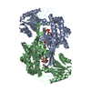
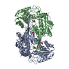
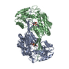
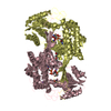
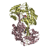
- PDB-4v3f: Crystal structure of betaine aldehyde dehydrogenase from spinach ... -
+ Open data

- Basic information
| Entry | Database: PDB / ID: 4v3f | ||||||
|---|---|---|---|---|---|---|---|
| Title | Crystal structure of betaine aldehyde dehydrogenase from spinach showing a thiohemiacetal with betaine aldehyde | ||||||
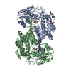
 Components Components | (BETAINE ALDEHYDE DEHYDROGENASE, ...) x 2 | ||||||
 Keywords Keywords | OXIDOREDUCTASE / BETAINE ALDEHYDE DEHYDROGENASE / ALDH10. | ||||||
| Function / homology |  Function and homology information Function and homology information3-aminopropanal dehydrogenase (NAD+) activity / 4-trimethylammoniobutyraldehyde dehydrogenase / 4-trimethylammoniobutyraldehyde dehydrogenase activity / aminobutyraldehyde dehydrogenase / aminobutyraldehyde dehydrogenase (NAD+) activity / betaine-aldehyde dehydrogenase (NAD+) activity / betaine-aldehyde dehydrogenase / : / Oxidoreductases; Acting on the aldehyde or oxo group of donors; With NAD+ or NADP+ as acceptor / aldehyde metabolic process ...3-aminopropanal dehydrogenase (NAD+) activity / 4-trimethylammoniobutyraldehyde dehydrogenase / 4-trimethylammoniobutyraldehyde dehydrogenase activity / aminobutyraldehyde dehydrogenase / aminobutyraldehyde dehydrogenase (NAD+) activity / betaine-aldehyde dehydrogenase (NAD+) activity / betaine-aldehyde dehydrogenase / : / Oxidoreductases; Acting on the aldehyde or oxo group of donors; With NAD+ or NADP+ as acceptor / aldehyde metabolic process / cellular detoxification of aldehyde / potassium ion binding / protein homodimerization activity Similarity search - Function | ||||||
| Biological species |  SPINACIA OLERACEA (spinach) SPINACIA OLERACEA (spinach) | ||||||
| Method |  X-RAY DIFFRACTION / SYNCHROTRON / MOLECULAR REPLACEMENT / Resolution: 1.998 Å X-RAY DIFFRACTION / SYNCHROTRON / MOLECULAR REPLACEMENT / Resolution: 1.998 Å | ||||||
 Authors Authors | Zarate-Romero, A. / Munoz-Clares, R.A. | ||||||
 Citation Citation | Journal: Biochem.J. / Year: 2016 Title: Reversible, Partial Inactivation of Plant Betaine Aldehyde Dehydrogenase by Betaine Aldehyde: Mechanism and Possible Physiological Implications. Authors: Zarate-Romero, A. / Murillo-Melo, D.S. / Mujica-Jimenez, C. / Montiel, C. / Munoz-Clares, R.A. | ||||||
| History |
|
- Structure visualization
| Structure viewer | Molecule: MolmilJmol/JSmol |
|---|
- Downloads & links
-Download
| PDBx/mmCIF format | 4v3f.cif.gz | 784.2 KB | Display | PDBx/mmCIF format |
|---|---|---|---|---|
| PDB format | pdb4v3f.ent.gz | 653.2 KB | Display | PDB format |
| PDBx/mmJSON format | 4v3f.json.gz | Tree view | PDBx/mmJSON format | |
| Others |  Other downloads Other downloads |
-Validation report
| Arichive directory | https://data.pdbj.org/pub/pdb/validation_reports/v3/4v3fftp://data.pdbj.org/pub/pdb/validation_reports/v3/4v3f | HTTPS FTP |
|---|
-Related structure data
| Related structure data |  5a2dC 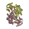 4a0mS S: Starting model for refinement C: citing same article ( |
|---|---|
| Similar structure data |

-Links
 PDBj
PDBj

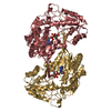
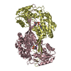
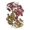
- Assembly
| Deposited unit | 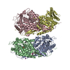
| ||||||||
|---|---|---|---|---|---|---|---|---|---|
| 1 | 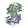
| ||||||||
| 2 | 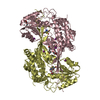
| ||||||||
| Unit cell |
|
-Components
-BETAINE ALDEHYDE DEHYDROGENASE, ... , 2 types, 4 molecules ABDC
| #1: Protein | Mass: 54343.027 Da / Num. of mol.: 3 Source method: isolated from a genetically manipulated source Source: (gene. exp.) SPINACIA OLERACEA (spinach) / Production host:  #2: Protein | | Mass: 54359.027 Da / Num. of mol.: 1 Source method: isolated from a genetically manipulated source Source: (gene. exp.) SPINACIA OLERACEA (spinach) / Production host: |
|---|
-Non-polymers , 6 types, 685 molecules 
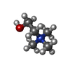


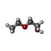

| #3: Chemical | ChemComp-IOD /  Mass: 126.904 Da / Num. of mol.: 15 / Source method: obtained synthetically / Formula: I Mass: 126.904 Da / Num. of mol.: 15 / Source method: obtained synthetically / Formula: I#4: Chemical |  Mass: 104.171 Da / Num. of mol.: 3 / Source method: obtained synthetically / Formula: C5H14NO Mass: 104.171 Da / Num. of mol.: 3 / Source method: obtained synthetically / Formula: C5H14NO#5: Chemical |  Mass: 194.226 Da / Num. of mol.: 3 / Source method: obtained synthetically / Formula: C8H18O5 / Comment: precipitant*YM Mass: 194.226 Da / Num. of mol.: 3 / Source method: obtained synthetically / Formula: C8H18O5 / Comment: precipitant*YM#6: Chemical |  Mass: 92.094 Da / Num. of mol.: 2 / Source method: obtained synthetically / Formula: C3H8O3 Mass: 92.094 Da / Num. of mol.: 2 / Source method: obtained synthetically / Formula: C3H8O3#7: Chemical | ChemComp-ETX / |  Mass: 90.121 Da / Num. of mol.: 1 / Source method: obtained synthetically / Formula: C4H10O2 Mass: 90.121 Da / Num. of mol.: 1 / Source method: obtained synthetically / Formula: C4H10O2#8: Water | ChemComp-HOH / | Mass: 18.015 Da / Num. of mol.: 661 / Source method: isolated from a natural source / Formula: H2O |
|---|
-Details
| Has protein modification | Y |
|---|
-Experimental details
-Experiment
| Experiment | Method: X-RAY DIFFRACTION / Number of used crystals: 1 |
|---|
- Sample preparation
| Crystal | Density Matthews: 2.15 Å3/Da / Density % sol: 42.7 % / Description: NONE |
|---|---|
| Crystal grow | pH: 6.2 Details: AMMONIUM IODIDE 0.2 M, POLY ETHYLENE GLYCOL 3350 20% (W/V), PH 6.2 |
-Data collection
| Diffraction | Mean temperature: 100 K |
|---|---|
| Diffraction source | Source: SYNCHROTRON / Site: APS  / Beamline: 19-BM / Wavelength: 0.9791 / Beamline: 19-BM / Wavelength: 0.9791 |
| Detector | Type: ADSC QUANTUM 210r / Detector: CCD / Date: Mar 13, 2014 |
| Radiation | Monochromator: SI (1 1 1) / Protocol: SINGLE WAVELENGTH / Monochromatic (M) / Laue (L): M / Scattering type: x-ray |
| Radiation wavelength | Wavelength: 0.9791 Å / Relative weight: 1 |
| Reflection | Resolution: 2→29 Å / Num. obs: 122131 / % possible obs: 96.1 % / Observed criterion σ(I): 2 / Redundancy: 2.4 % / Rmerge(I) obs: 0.048 / Rsym value: 0.048 / Net I/σ(I): 9.1 |
| Reflection shell | Resolution: 2→2.11 Å / Redundancy: 1.7 % / Rmerge(I) obs: 0.268 / Mean I/σ(I) obs: 2.3 / % possible all: 88 |
- Processing
| Software |
| |||||||||||||||||||||||||||||||||||||||||||||||||||||||||||||||||||||||||||||||||||||||||||||||||||||||||||||||||||||||||||||||||||||||||||||||||||||||||||||||||||||||||||||||||||||||||||||||||||||||||||||||||||||||||
|---|---|---|---|---|---|---|---|---|---|---|---|---|---|---|---|---|---|---|---|---|---|---|---|---|---|---|---|---|---|---|---|---|---|---|---|---|---|---|---|---|---|---|---|---|---|---|---|---|---|---|---|---|---|---|---|---|---|---|---|---|---|---|---|---|---|---|---|---|---|---|---|---|---|---|---|---|---|---|---|---|---|---|---|---|---|---|---|---|---|---|---|---|---|---|---|---|---|---|---|---|---|---|---|---|---|---|---|---|---|---|---|---|---|---|---|---|---|---|---|---|---|---|---|---|---|---|---|---|---|---|---|---|---|---|---|---|---|---|---|---|---|---|---|---|---|---|---|---|---|---|---|---|---|---|---|---|---|---|---|---|---|---|---|---|---|---|---|---|---|---|---|---|---|---|---|---|---|---|---|---|---|---|---|---|---|---|---|---|---|---|---|---|---|---|---|---|---|---|---|---|---|---|---|---|---|---|---|---|---|---|---|---|---|---|---|---|---|---|
| Refinement | Method to determine structure: MOLECULAR REPLACEMENT Starting model: PDB ENTRY 4A0M Resolution: 1.998→28.968 Å / SU ML: 0.23 / σ(F): 1.96 / Phase error: 23.07 / Stereochemistry target values: ML
| |||||||||||||||||||||||||||||||||||||||||||||||||||||||||||||||||||||||||||||||||||||||||||||||||||||||||||||||||||||||||||||||||||||||||||||||||||||||||||||||||||||||||||||||||||||||||||||||||||||||||||||||||||||||||
| Solvent computation | Shrinkage radii: 0.9 Å / VDW probe radii: 1.11 Å / Solvent model: FLAT BULK SOLVENT MODEL | |||||||||||||||||||||||||||||||||||||||||||||||||||||||||||||||||||||||||||||||||||||||||||||||||||||||||||||||||||||||||||||||||||||||||||||||||||||||||||||||||||||||||||||||||||||||||||||||||||||||||||||||||||||||||
| Refinement step | Cycle: LAST / Resolution: 1.998→28.968 Å
| |||||||||||||||||||||||||||||||||||||||||||||||||||||||||||||||||||||||||||||||||||||||||||||||||||||||||||||||||||||||||||||||||||||||||||||||||||||||||||||||||||||||||||||||||||||||||||||||||||||||||||||||||||||||||
| Refine LS restraints |
| |||||||||||||||||||||||||||||||||||||||||||||||||||||||||||||||||||||||||||||||||||||||||||||||||||||||||||||||||||||||||||||||||||||||||||||||||||||||||||||||||||||||||||||||||||||||||||||||||||||||||||||||||||||||||
| LS refinement shell |
|
