 Movie
Movie Controller


+ Open data

- Basic information
| Entry | Database: PDB / ID: 5ljn | ||||||
|---|---|---|---|---|---|---|---|
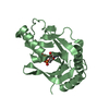
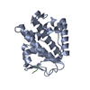
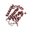
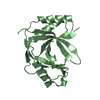
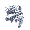
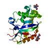
| Title | Structure of the HOIP PUB domain bound to SPATA2 PIM peptide | ||||||
 Components Components |
| ||||||
 Keywords Keywords | LIGASE / PUB domain | ||||||
| Function / homology |  Function and homology information Function and homology informationprotein linear deubiquitination / protein linear polyubiquitination / LUBAC complex / linear polyubiquitin binding / RBR-type E3 ubiquitin transferase / CD40 signaling pathway / positive regulation of xenophagy / regulation of necroptotic process / CD40 receptor complex / programmed cell death ...protein linear deubiquitination / protein linear polyubiquitination / LUBAC complex / linear polyubiquitin binding / RBR-type E3 ubiquitin transferase / CD40 signaling pathway / positive regulation of xenophagy / regulation of necroptotic process / CD40 receptor complex / programmed cell death / regulation of tumor necrosis factor-mediated signaling pathway / TNFR1-induced proapoptotic signaling / : / K48-linked polyubiquitin modification-dependent protein binding / protein K63-linked deubiquitination / K63-linked polyubiquitin modification-dependent protein binding / ubiquitin-specific protease binding / TNFR1-induced NF-kappa-B signaling pathway / ubiquitin binding / Regulation of TNFR1 signaling / cytoplasmic side of plasma membrane / protein polyubiquitination / ubiquitin-protein transferase activity / ubiquitin protein ligase activity / T cell receptor signaling pathway / signaling receptor complex adaptor activity / regulation of inflammatory response / spermatogenesis / positive regulation of canonical NF-kappaB signal transduction / defense response to bacterium / ubiquitin protein ligase binding / protein-containing complex binding / zinc ion binding / identical protein binding / nucleus / cytoplasm / cytosol Similarity search - Function | ||||||
| Biological species |  Homo sapiens (human) Homo sapiens (human) | ||||||
| Method |  X-RAY DIFFRACTION / SYNCHROTRON / MOLECULAR REPLACEMENT / Resolution: 2.701 Å X-RAY DIFFRACTION / SYNCHROTRON / MOLECULAR REPLACEMENT / Resolution: 2.701 Å | ||||||
 Authors Authors | Elliott, P.R. / Komander, D. | ||||||
 Citation Citation | Journal: Mol.Cell / Year: 2016 Title: SPATA2 Links CYLD to LUBAC, Activates CYLD, and Controls LUBAC Signaling. Authors: Elliott, P.R. / Leske, D. / Hrdinka, M. / Bagola, K. / Fiil, B.K. / McLaughlin, S.H. / Wagstaff, J. / Volkmar, N. / Christianson, J.C. / Kessler, B.M. / Freund, S.M. / Komander, D. / Gyrd-Hansen, M. | ||||||
| History |
|
- Structure visualization
| Structure viewer | Molecule: MolmilJmol/JSmol |
|---|
- Downloads & links
-Download
| PDBx/mmCIF format | 5ljn.cif.gz | 80.4 KB | Display | PDBx/mmCIF format |
|---|---|---|---|---|
| PDB format | pdb5ljn.ent.gz | 59.5 KB | Display | PDB format |
| PDBx/mmJSON format | 5ljn.json.gz | Tree view | PDBx/mmJSON format | |
| Others |  Other downloads Other downloads |
-Validation report
| Arichive directory | https://data.pdbj.org/pub/pdb/validation_reports/lj/5ljnftp://data.pdbj.org/pub/pdb/validation_reports/lj/5ljn | HTTPS FTP |
|---|
-Related structure data
| Related structure data | 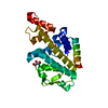 5ljmC  4oykS S: Starting model for refinement C: citing same article ( |
|---|---|
| Similar structure data |


-Links
 PDBj
PDBj


- Assembly
| Deposited unit | 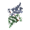
| ||||||||
|---|---|---|---|---|---|---|---|---|---|
| 1 | 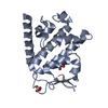
| ||||||||
| 2 | 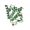
| ||||||||
| Unit cell |
|
-Components
| #1: Protein | Mass: 19647.059 Da / Num. of mol.: 2 Source method: isolated from a genetically manipulated source Source: (gene. exp.) Homo sapiens (human) / Gene: RNF31, ZIBRA / Production host:  References: UniProt: Q96EP0, Ligases; Forming carbon-nitrogen bonds; Acid-amino-acid ligases (peptide synthases) #2: Protein/peptide | Mass: 926.923 Da / Num. of mol.: 2 / Fragment: UNP residues 334-341 / Source method: obtained synthetically / Source: (synth.) Homo sapiens (human) / References: UniProt: Q9UM82#3: Chemical |   Mass: 96.063 Da / Num. of mol.: 3 / Source method: obtained synthetically / Formula: SO4 Mass: 96.063 Da / Num. of mol.: 3 / Source method: obtained synthetically / Formula: SO4#4: Chemical | ChemComp-GOL / |   Mass: 92.094 Da / Num. of mol.: 1 / Source method: obtained synthetically / Formula: C3H8O3 Mass: 92.094 Da / Num. of mol.: 1 / Source method: obtained synthetically / Formula: C3H8O3Has protein modification | Y | |
|---|
-Experimental details
-Experiment
| Experiment | Method: X-RAY DIFFRACTION / Number of used crystals: 1 |
|---|
- Sample preparation
| Crystal | Density Matthews: 2.69 Å3/Da / Density % sol: 54.31 % |
|---|---|
| Crystal grow | Temperature: 293 K / Method: vapor diffusion, sitting drop Details: 1.7-1.9 M (NH4)2SO4, 50 mM sodium cacodylate pH 6.4-7.0, 15 mM MgCl2 Protein:precipitant ratio; 1:2 200 nl drops PH range: 6.4-7.0 |
-Data collection
| Diffraction | Mean temperature: 100 K | ||||||||||||||||||
|---|---|---|---|---|---|---|---|---|---|---|---|---|---|---|---|---|---|---|---|
| Diffraction source | Source: SYNCHROTRON / Site: Diamond  / Beamline: I02 / Wavelength: 0.9795 Å / Beamline: I02 / Wavelength: 0.9795 Å | ||||||||||||||||||
| Detector | Type: DECTRIS PILATUS3 6M / Detector: PIXEL / Date: Oct 10, 2015 | ||||||||||||||||||
| Radiation | Protocol: SINGLE WAVELENGTH / Monochromatic (M) / Laue (L): M / Scattering type: x-ray | ||||||||||||||||||
| Radiation wavelength | Wavelength: 0.9795 Å / Relative weight: 1 | ||||||||||||||||||
| Reflection | Resolution: 2.7→62.96 Å / Num. obs: 11439 / % possible obs: 97.7 % / Redundancy: 2.7 % / Biso Wilson estimate: 57.09 Å2 / CC1/2: 0.997 / Rmerge(I) obs: 0.08 / Rpim(I) all: 0.057 / Rrim(I) all: 0.098 / Net I/σ(I): 9.5 / Num. measured all: 31067 | ||||||||||||||||||
| Reflection shell |
|
- Processing
| Software |
| |||||||||||||||||||||||||||||||||||
|---|---|---|---|---|---|---|---|---|---|---|---|---|---|---|---|---|---|---|---|---|---|---|---|---|---|---|---|---|---|---|---|---|---|---|---|---|
| Refinement | Method to determine structure: MOLECULAR REPLACEMENT Starting model: 4OYK Resolution: 2.701→62.96 Å / SU ML: 0.42 / Cross valid method: FREE R-VALUE / σ(F): 1.35 / Phase error: 33.28
| |||||||||||||||||||||||||||||||||||
| Solvent computation | Shrinkage radii: 0.9 Å / VDW probe radii: 1.11 Å | |||||||||||||||||||||||||||||||||||
| Displacement parameters | Biso max: 122.7 Å2 / Biso mean: 61.8122 Å2 / Biso min: 29.86 Å2 | |||||||||||||||||||||||||||||||||||
| Refinement step | Cycle: final / Resolution: 2.701→62.96 Å
| |||||||||||||||||||||||||||||||||||
| Refine LS restraints |
| |||||||||||||||||||||||||||||||||||
| LS refinement shell | Refine-ID: X-RAY DIFFRACTION / Total num. of bins used: 4
|
