 Movie
Movie Controller


+ Open data

- Basic information
| Entry | Database: PDB / ID: 5ldk | ||||||
|---|---|---|---|---|---|---|---|
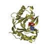
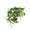
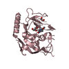
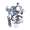
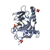
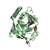
| Title | Crystal structure of E.coli LigT complexed with ATP | ||||||
 Components Components | RNA 2',3'-cyclic phosphodiesterase | ||||||
 Keywords Keywords | HYDROLASE / Enzyme / 2H phosphoesterase/ligase | ||||||
| Function / homology |  Function and homology information Function and homology informationRNA 2',3'-cyclic 3'-phosphodiesterase / RNA 2',3'-cyclic 3'-phosphodiesterase activity / 2',3'-cyclic-nucleotide 3'-phosphodiesterase activity / ligase activity / ATP binding Similarity search - Function | ||||||
| Biological species |  | ||||||
| Method |  X-RAY DIFFRACTION / SYNCHROTRON / MOLECULAR REPLACEMENT / Resolution: 2.099 Å X-RAY DIFFRACTION / SYNCHROTRON / MOLECULAR REPLACEMENT / Resolution: 2.099 Å | ||||||
 Authors Authors | Myllykoski, M. / Kursula, P. | ||||||
 Citation Citation | Journal: PLoS ONE / Year: 2017 Title: Structural aspects of nucleotide ligand binding by a bacterial 2H phosphoesterase. Authors: Myllykoski, M. / Kursula, P. | ||||||
| History |
|
- Structure visualization
| Structure viewer | Molecule: MolmilJmol/JSmol |
|---|
- Downloads & links
-Download
| PDBx/mmCIF format | 5ldk.cif.gz | 85.6 KB | Display | PDBx/mmCIF format |
|---|---|---|---|---|
| PDB format | pdb5ldk.ent.gz | 64.2 KB | Display | PDB format |
| PDBx/mmJSON format | 5ldk.json.gz | Tree view | PDBx/mmJSON format | |
| Others |  Other downloads Other downloads |
-Validation report
| Arichive directory | https://data.pdbj.org/pub/pdb/validation_reports/ld/5ldkftp://data.pdbj.org/pub/pdb/validation_reports/ld/5ldk | HTTPS FTP |
|---|
-Related structure data
| Related structure data |  5ldiSC 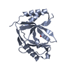 5ldjC  5ldmC 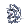 5ldoC  5ldpC  5ldqC S: Starting model for refinement C: citing same article ( |
|---|---|
| Similar structure data |


-Links
 PDBj
PDBj- Assembly
| Deposited unit | 
| ||||||||
|---|---|---|---|---|---|---|---|---|---|
| 1 | 
| ||||||||
| 2 | 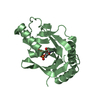
| ||||||||
| Unit cell |
|
-Components
| #1: Protein | Mass: 20045.029 Da / Num. of mol.: 2 Source method: isolated from a genetically manipulated source Source: (gene. exp.) Gene: thpR, ECBD_3472 / Plasmid: pTH 27 / Production host: References: UniProt: A0A140NFI1, Hydrolases; Acting on ester bonds; Phosphoric-diester hydrolases #2: Chemical | ChemComp-ATP / |   Mass: 507.181 Da / Num. of mol.: 1 / Source method: obtained synthetically / Formula: C10H16N5O13P3 / Comment: ATP, energy-carrying molecule*YM Mass: 507.181 Da / Num. of mol.: 1 / Source method: obtained synthetically / Formula: C10H16N5O13P3 / Comment: ATP, energy-carrying molecule*YM#3: Water | ChemComp-HOH / |  Mass: 18.015 Da / Num. of mol.: 197 / Source method: isolated from a natural source / Formula: H2O Mass: 18.015 Da / Num. of mol.: 197 / Source method: isolated from a natural source / Formula: H2O |
|---|
-Experimental details
-Experiment
| Experiment | Method: X-RAY DIFFRACTION / Number of used crystals: 1 |
|---|
- Sample preparation
| Crystal | Density Matthews: 2.83 Å3/Da / Density % sol: 56.47 % |
|---|---|
| Crystal grow | Temperature: 293 K / Method: vapor diffusion, sitting drop / pH: 7.5 Details: 0.1 M Tris-HCl pH 7.5, 0.2 M MgCl2, 18% (w/v) PEG 8000 |
-Data collection
| Diffraction | Mean temperature: 100 K |
|---|---|
| Diffraction source | Source: SYNCHROTRON / Site: MAX II  / Beamline: I911-2 / Wavelength: 1.04 Å / Beamline: I911-2 / Wavelength: 1.04 Å |
| Detector | Type: MAR CCD 165 mm / Detector: CCD / Date: Jun 12, 2013 |
| Radiation | Protocol: SINGLE WAVELENGTH / Monochromatic (M) / Laue (L): M / Scattering type: x-ray |
| Radiation wavelength | Wavelength: 1.04 Å / Relative weight: 1 |
| Reflection | Resolution: 2.099→30 Å / Num. obs: 26124 / % possible obs: 99.2 % / Redundancy: 3.81 % / Biso Wilson estimate: 32 Å2 / CC1/2: 0.998 / Rmerge(I) obs: 0.075 / Net I/σ(I): 14.16 |
| Reflection shell | Resolution: 2.1→2.15 Å / Redundancy: 3.78 % / Rmerge(I) obs: 0.829 / Mean I/σ(I) obs: 1.65 / % possible all: 99.2 |
- Processing
| Software |
| |||||||||||||||||||||||||||||||||||||||||||||||||||||||||||||||||||||||||||||||||||||||||||||||||||||||||
|---|---|---|---|---|---|---|---|---|---|---|---|---|---|---|---|---|---|---|---|---|---|---|---|---|---|---|---|---|---|---|---|---|---|---|---|---|---|---|---|---|---|---|---|---|---|---|---|---|---|---|---|---|---|---|---|---|---|---|---|---|---|---|---|---|---|---|---|---|---|---|---|---|---|---|---|---|---|---|---|---|---|---|---|---|---|---|---|---|---|---|---|---|---|---|---|---|---|---|---|---|---|---|---|---|---|---|
| Refinement | Method to determine structure: MOLECULAR REPLACEMENT Starting model: PDBid: 5ldi Resolution: 2.099→23.37 Å / SU ML: 0.26 / Cross valid method: FREE R-VALUE / σ(F): 2.01 / Phase error: 24.92
| |||||||||||||||||||||||||||||||||||||||||||||||||||||||||||||||||||||||||||||||||||||||||||||||||||||||||
| Solvent computation | Shrinkage radii: 0.9 Å / VDW probe radii: 1.11 Å | |||||||||||||||||||||||||||||||||||||||||||||||||||||||||||||||||||||||||||||||||||||||||||||||||||||||||
| Displacement parameters | Biso mean: 36.45 Å2 | |||||||||||||||||||||||||||||||||||||||||||||||||||||||||||||||||||||||||||||||||||||||||||||||||||||||||
| Refinement step | Cycle: LAST / Resolution: 2.099→23.37 Å
| |||||||||||||||||||||||||||||||||||||||||||||||||||||||||||||||||||||||||||||||||||||||||||||||||||||||||
| Refine LS restraints |
| |||||||||||||||||||||||||||||||||||||||||||||||||||||||||||||||||||||||||||||||||||||||||||||||||||||||||
| LS refinement shell |
|
