 Movie
Movie Controller


+ Open data

- Basic information
| Entry | Database: PDB / ID: 5zbo | ||||||
|---|---|---|---|---|---|---|---|
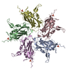
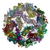
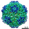
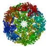
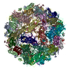
| Title | Cryo-EM structure of PCV2 VLPs | ||||||
 Components Components | Capsid protein | ||||||
 Keywords Keywords | DNA BINDING PROTEIN / Porcine circle virus type 2 (PCV2) / capsid protein / virus-like-particle (VLP) / type-specific epitope | ||||||
| Function / homology |  Function and homology information Function and homology informationviral capsid assembly / T=1 icosahedral viral capsid / viral penetration into host nucleus / host cell / endocytosis involved in viral entry into host cell / virion attachment to host cell / host cell nucleus / DNA binding Similarity search - Function | ||||||
| Biological species |  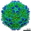 Porcine circovirus 2 Porcine circovirus 2 | ||||||
| Method | ELECTRON MICROSCOPY / single particle reconstruction / cryo EM / Resolution: 4.12 Å | ||||||
 Authors Authors | Mo, X. / Yuan, A.Y. | ||||||
 Citation Citation | Journal: PLoS Pathog / Year: 2019 Title: Structural roles of PCV2 capsid protein N-terminus in PCV2 particle assembly and identification of PCV2 type-specific neutralizing epitope. Authors: Xiaobing Mo / Xiangdong Li / Bo Yin / Junhua Deng / Kegong Tian / Adam Yuan /   Abstract: Postweaning multisystemic wasting disease (PMWS) in piglets caused by porcine circovirus type 2 (PCV2) is one of the major threats to most pig farms worldwide. Among all the PCV types, PCV2 is the ...Postweaning multisystemic wasting disease (PMWS) in piglets caused by porcine circovirus type 2 (PCV2) is one of the major threats to most pig farms worldwide. Among all the PCV types, PCV2 is the dominant genotype causing PMWS and associated diseases. Considerable efforts were made to study the virus-like-particle (VLP) assembly and the specific PCV2-associated epitope(s) in order to establish the solid foundation for engineered PCV2 vaccine development. Although the N-terminal fragment including Nuclear Localization Signal (NLS) sequence seems important for recombinant PCV2 capsid protein expression and VLP assembly, the detailed structural and functional information regarding this important fragment are largely unknown. In this study, we report crystal structure of PCV2 VLP assembled from N-terminal NLS truncated PCV2 capsid protein at 2.8 Å resolution and cryo-EM structure of PCV2 VLP assembled from full-length PCV2 capsid protein at 4.1Å resolution. Our in vitro PCV2 VLP assembly results show that NLS-truncated PCV2 capsid protein only forms instable VLPs which were easily disassembled in solution, whereas full-length PCV2 capsid protein forms stable VLPs due to interaction between 15PRSHLGQILRRRP27 (α-helix) and 33RHRYRWRRKN42 (NLS-B) in a repeated manner. In addition, our results also showed that N-terminal truncation of PCV2 capsid protein up to 27 residues still forms PCV2 particles in solution with similar size and immunogenicity, while N-terminal truncation of PCV2 capsid protein with more than 30 residues is not able to form stable PCV2 particles in solution, demonstrating the importance of interaction between the α-helix at N-terminal and NLS-B in PCV2 VLP formation. Moreover, we also report the cryo-EM structure of PCV2 VLP in complex with 3H11-Fab, a PCV2 type-specific neutralizing antibody, at 15 Å resolution. MAb-3H11 specifically recognizes one exposed epitope located on the VLP surface EF-loop (residues 128-143), which is further confirmed by PCV1-PCV2 epitope swapping assay. Hence, our results have revealed the structural roles of N-terminal fragment of PCV2 capsid protein in PCV2 particle assembly and pinpointed one PCV2 type-specific neutralizing epitope for the first time, which could provide clear clue for next generation PCV2 vaccine and diagnostic kits development. | ||||||
| History |
|
- Structure visualization
| Movie |
Movie viewer |
|---|---|
| Structure viewer | Molecule: MolmilJmol/JSmol |
- Downloads & links
-Download
| PDBx/mmCIF format | 5zbo.cif.gz | 1.8 MB | Display | PDBx/mmCIF format |
|---|---|---|---|---|
| PDB format | pdb5zbo.ent.gz | 1.6 MB | Display | PDB format |
| PDBx/mmJSON format | 5zbo.json.gz | Tree view | PDBx/mmJSON format | |
| Others |  Other downloads Other downloads |
-Validation report
| Arichive directory | https://data.pdbj.org/pub/pdb/validation_reports/zb/5zboftp://data.pdbj.org/pub/pdb/validation_reports/zb/5zbo | HTTPS FTP |
|---|
-Related structure data
| Related structure data |  6746MC 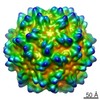 6961C  5zjuC M: map data used to model this data C: citing same article ( |
|---|---|
| Similar structure data |


-Links
 PDBj
PDBj
- Assembly
| Deposited unit | 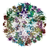
|
|---|---|
| 1 |
|
-Components
| #1: Protein | Mass: 24126.051 Da / Num. of mol.: 60 Source method: isolated from a genetically manipulated source Source: (gene. exp.) Porcine circovirus 2 / Gene: cap / Production host:  |
|---|
-Experimental details
-Experiment
| Experiment | Method: ELECTRON MICROSCOPY |
|---|---|
| EM experiment | Aggregation state: PARTICLE / 3D reconstruction method: single particle reconstruction |
- Sample preparation
| Component | Name: Porcine circovirus 2 cap / Type: VIRUS / Entity ID: all / Source: RECOMBINANT |
|---|---|
| Source (natural) | Organism: Porcine circovirus 2 |
| Source (recombinant) | Organism: |
| Details of virus | Empty: YES / Enveloped: NO / Isolate: OTHER / Type: VIRUS-LIKE PARTICLE |
| Buffer solution | pH: 6.5 |
| Specimen | Embedding applied: NO / Shadowing applied: NO / Staining applied: NO / Vitrification applied: YES |
| Vitrification | Cryogen name: NITROGEN |
- Electron microscopy imaging
| Experimental equipment |  Model: Titan Krios / Image courtesy: FEI Company |
|---|---|
| Microscopy | Model: FEI TITAN KRIOS |
| Electron gun | Electron source:  FIELD EMISSION GUN / Accelerating voltage: 300 kV / Illumination mode: OTHER FIELD EMISSION GUN / Accelerating voltage: 300 kV / Illumination mode: OTHER |
| Electron lens | Mode: DARK FIELD |
| Image recording | Electron dose: 25 e/Å2 / Film or detector model: FEI FALCON I (4k x 4k) |
- Processing
| Software | Name: PHENIX / Version: (1.14_3260: phenix.real_space_refine) / Classification: refinement | ||||||||||||||||||||||||
|---|---|---|---|---|---|---|---|---|---|---|---|---|---|---|---|---|---|---|---|---|---|---|---|---|---|
| EM software |
| ||||||||||||||||||||||||
| CTF correction | Type: PHASE FLIPPING AND AMPLITUDE CORRECTION | ||||||||||||||||||||||||
| 3D reconstruction | Resolution: 4.12 Å / Resolution method: FSC 0.143 CUT-OFF / Num. of particles: 1200 / Symmetry type: POINT | ||||||||||||||||||||||||
| Atomic model building | Space: REAL | ||||||||||||||||||||||||
| Refinement | Stereochemistry target values: CDL v1.2 | ||||||||||||||||||||||||
| Refine LS restraints |
|
