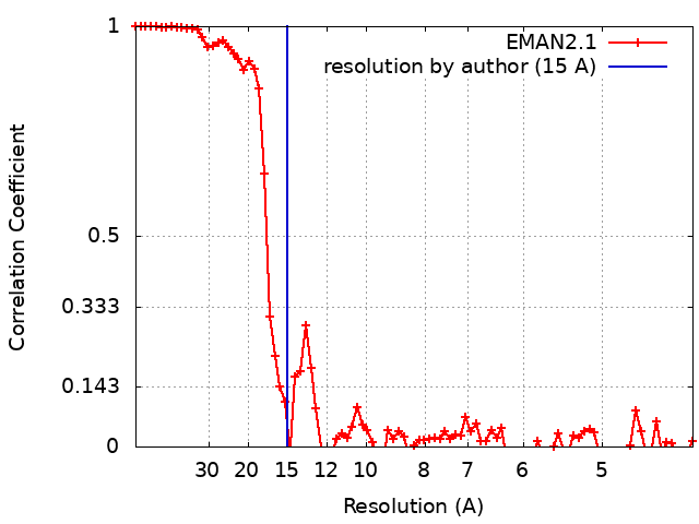
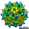
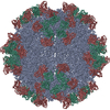
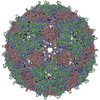
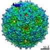
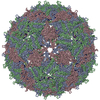
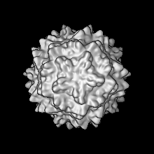
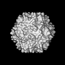
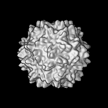
Journal: PLoS Pathog / Year: 2019 Title: Structural roles of PCV2 capsid protein N-terminus in PCV2 particle assembly and identification of PCV2 type-specific neutralizing epitope. Authors: Xiaobing Mo / Xiangdong Li / Bo Yin / Junhua Deng / Kegong Tian / Adam Yuan / Abstract: Postweaning multisystemic wasting disease (PMWS) in piglets caused by porcine circovirus type 2 (PCV2) is one of the major threats to most pig farms worldwide. Among all the PCV types, PCV2 is the ...Postweaning multisystemic wasting disease (PMWS) in piglets caused by porcine circovirus type 2 (PCV2) is one of the major threats to most pig farms worldwide. Among all the PCV types, PCV2 is the dominant genotype causing PMWS and associated diseases. Considerable efforts were made to study the virus-like-particle (VLP) assembly and the specific PCV2-associated epitope(s) in order to establish the solid foundation for engineered PCV2 vaccine development. Although the N-terminal fragment including Nuclear Localization Signal (NLS) sequence seems important for recombinant PCV2 capsid protein expression and VLP assembly, the detailed structural and functional information regarding this important fragment are largely unknown. In this study, we report crystal structure of PCV2 VLP assembled from N-terminal NLS truncated PCV2 capsid protein at 2.8 Å resolution and cryo-EM structure of PCV2 VLP assembled from full-length PCV2 capsid protein at 4.1Å resolution. Our in vitro PCV2 VLP assembly results show that NLS-truncated PCV2 capsid protein only forms instable VLPs which were easily disassembled in solution, whereas full-length PCV2 capsid protein forms stable VLPs due to interaction between 15PRSHLGQILRRRP27 (α-helix) and 33RHRYRWRRKN42 (NLS-B) in a repeated manner. In addition, our results also showed that N-terminal truncation of PCV2 capsid protein up to 27 residues still forms PCV2 particles in solution with similar size and immunogenicity, while N-terminal truncation of PCV2 capsid protein with more than 30 residues is not able to form stable PCV2 particles in solution, demonstrating the importance of interaction between the α-helix at N-terminal and NLS-B in PCV2 VLP formation. Moreover, we also report the cryo-EM structure of PCV2 VLP in complex with 3H11-Fab, a PCV2 type-specific neutralizing antibody, at 15 Å resolution. MAb-3H11 specifically recognizes one exposed epitope located on the VLP surface EF-loop (residues 128-143), which is further confirmed by PCV1-PCV2 epitope swapping assay. Hence, our results have revealed the structural roles of N-terminal fragment of PCV2 capsid protein in PCV2 particle assembly and pinpointed one PCV2 type-specific neutralizing epitope for the first time, which could provide clear clue for next generation PCV2 vaccine and diagnostic kits development.
History
Deposition
May 3, 2018
-
Header (metadata) release
May 16, 2018
-
Map release
May 16, 2018
-
Update
Mar 13, 2019
-
Current status
Mar 13, 2019
Processing site: PDBj / Status: Released
-
Structure visualization
Movie
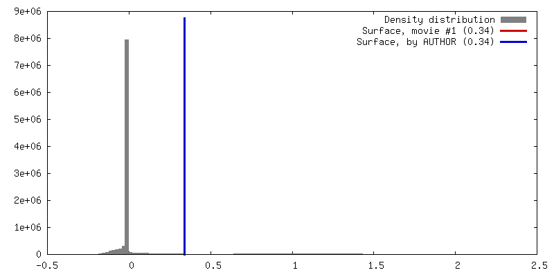
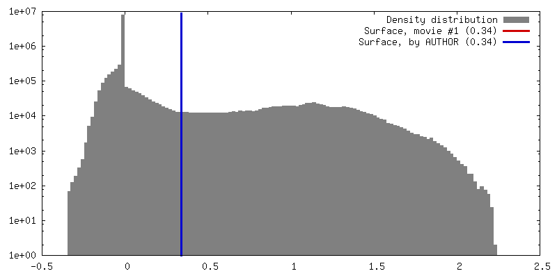
Surface view with section colored by density value
Model: Quantifoil R2/2 / Material: COPPER / Mesh: 400 / Support film - Material: FORMVAR / Support film - topology: HOLEY / Pretreatment - Type: GLOW DISCHARGE / Pretreatment - Atmosphere: AIR / Pretreatment - Pressure: 101.325 kPa
Vitrification
Cryogen name: ETHANE / Chamber humidity: 100 % / Chamber temperature: 298 K / Instrument: FEI VITROBOT MARK IV / Details: blot for 5 seconds before pluning.
Details
This sample was monodisperse
-
Electron microscopy
Microscope
FEI TITAN
Temperature
Min: 70.0 K / Max: 70.0 K
Details
Preliminary grid screening was performed manually.
Image recording
Film or detector model: FEI FALCON II (4k x 4k) / Number grids imaged: 5 / Number real images: 99 / Average exposure time: 1.0 sec. / Average electron dose: 35.0 e/Å2
Electron beam
Acceleration voltage: 300 kV / Electron source: FIELD EMISSION GUN
In the structure databanks used in Yorodumi, some data are registered as the other names, "COVID-19 virus" and "2019-nCoV". Here are the details of the virus and the list of structure data.
Jan 31, 2019. EMDB accession codes are about to change! (news from PDBe EMDB page)
EMDB accession codes are about to change! (news from PDBe EMDB page)
The allocation of 4 digits for EMDB accession codes will soon come to an end. Whilst these codes will remain in use, new EMDB accession codes will include an additional digit and will expand incrementally as the available range of codes is exhausted. The current 4-digit format prefixed with “EMD-” (i.e. EMD-XXXX) will advance to a 5-digit format (i.e. EMD-XXXXX), and so on. It is currently estimated that the 4-digit codes will be depleted around Spring 2019, at which point the 5-digit format will come into force.
The EM Navigator/Yorodumi systems omit the EMD- prefix.
Related info.:Q: What is EMD? / ID/Accession-code notation in Yorodumi/EM Navigator
Yorodumi is a browser for structure data from EMDB, PDB, SASBDB, etc.
This page is also the successor to EM Navigator detail page, and also detail information page/front-end page for Omokage search.
The word "yorodu" (or yorozu) is an old Japanese word meaning "ten thousand". "mi" (miru) is to see.
Related info.:EMDB / PDB / SASBDB / Comparison of 3 databanks / Yorodumi Search / Aug 31, 2016. New EM Navigator & Yorodumi / Yorodumi Papers / Jmol/JSmol / Function and homology information / Changes in new EM Navigator and Yorodumi
 Movie
Movie Controller


 Open data
 Basic information
 Map data
 Sample
 Function and homology information
 Porcine circovirus 2
 Authors
 Citation

 Structure visualization
 Downloads & links
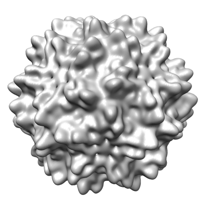 emd_6961.png
 http://ftp.pdbj.org/pub/emdb/structures/EMD-6961


 Z (Sec.)
 Y (Row.)
 X (Col.)


 Sample components
 Processing
 Electron microscopy
 FIELD EMISSION GUN