 Movie
Movie Controller


+ Open data

- Basic information
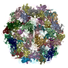
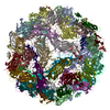
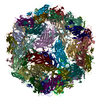
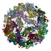
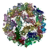
| Entry | Database: PDB / ID: 6ola | ||||||||||||
|---|---|---|---|---|---|---|---|---|---|---|---|---|---|
| Title | Structure of the PCV2d virus-like particle | ||||||||||||
 Components Components |
| ||||||||||||
 Keywords Keywords | VIRUS LIKE PARTICLE / Circovirus / viral jelly roll | ||||||||||||
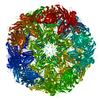
| Function / homology |  Function and homology information Function and homology informationviral capsid assembly / T=1 icosahedral viral capsid / viral penetration into host nucleus / host cell / endocytosis involved in viral entry into host cell / virion attachment to host cell / host cell nucleus / DNA binding Similarity search - Function | ||||||||||||
| Biological species |  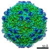 Porcine circovirus 2 Porcine circovirus 2 | ||||||||||||
| Method | ELECTRON MICROSCOPY / single particle reconstruction / cryo EM / Resolution: 3.3 Å | ||||||||||||
 Authors Authors | Khayat, R. / Wen, K. / Alimova, A. / Galarza, J. / Gottlieb, P. | ||||||||||||
| Funding support |  United States, 3items United States, 3items
| ||||||||||||
 Citation Citation | Journal: Virology / Year: 2019 Title: Structural characterization of the PCV2d virus-like particle at 3.3 Å resolution reveals differences to PCV2a and PCV2b capsids, a tetranucleotide, and an N-terminus near the icosahedral 3-fold axes. Authors: Reza Khayat / Ke Wen / Aleksandra Alimova / Boris Gavrilov / Al Katz / Jose M Galarza / Paul Gottlieb / Abstract: Porcine circovirus 2 (PCV2) has a major impact on the swine industry. Eight PCV2 genotypes (a-h) have been identified using capsid sequence analysis. PCV2d has been designated as the emerging ...Porcine circovirus 2 (PCV2) has a major impact on the swine industry. Eight PCV2 genotypes (a-h) have been identified using capsid sequence analysis. PCV2d has been designated as the emerging genotype. The cryo-electron microscopy molecular envelope of PCV2d virus-like particles identifies differences between PCV2a, b and d genotypes that accompany the emergence of PCV2b from PCV2a, and PCV2d from PCV2b. These differences indicate that sequence analysis of genotypes is insufficient, and that it is important to determine the PCV2 capsid structure as the virus evolves. Structure-based sequence comparison demonstrate that each genotype possesses a unique combination of amino acids located on the surface of the capsid that undergo substitution. We also demonstrate that the capsid N-terminus moves in response to increasing amount of nucleic acid packaged into the capsid. Furthermore, we model a tetranucleotide between the 5- and 2-fold axes of symmetry that appears to be responsible for capsid stability. | ||||||||||||
| History |
|
- Structure visualization
| Movie |
Movie viewer |
|---|---|
| Structure viewer | Molecule: MolmilJmol/JSmol |
- Downloads & links
-Download
| PDBx/mmCIF format | 6ola.cif.gz | 3.9 MB | Display | PDBx/mmCIF format |
|---|---|---|---|---|
| PDB format | pdb6ola.ent.gz | Display | PDB format | |
| PDBx/mmJSON format | 6ola.json.gz | Tree view | PDBx/mmJSON format | |
| Others |  Other downloads Other downloads |
-Validation report
| Arichive directory | https://data.pdbj.org/pub/pdb/validation_reports/ol/6olaftp://data.pdbj.org/pub/pdb/validation_reports/ol/6ola | HTTPS FTP |
|---|
-Related structure data
| Related structure data |  20113MC M: map data used to model this data C: citing same article ( |
|---|---|
| Similar structure data |


-Links
 PDBj
PDBj
- Assembly
| Deposited unit | 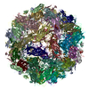
|
|---|---|
| 1 |
|
-Components
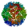
| #1: Protein | Mass: 23070.059 Da / Num. of mol.: 60 / Fragment: UNP residues 36-231 Source method: isolated from a genetically manipulated source Source: (gene. exp.) Porcine circovirus 2 / Gene: ORF2 / Cell line (production host): HEK293 / Production host:  Homo sapiens (human) / References: UniProt: G0ZPI6 Homo sapiens (human) / References: UniProt: G0ZPI6#2: DNA chain | Mass: 1191.818 Da / Num. of mol.: 60 Source method: isolated from a genetically manipulated source Source: (gene. exp.) Porcine circovirus 2 / Cell line (production host): HEK293 / Production host: Homo sapiens (human) |
|---|
-Experimental details
-Experiment
| Experiment | Method: ELECTRON MICROSCOPY |
|---|---|
| EM experiment | Aggregation state: PARTICLE / 3D reconstruction method: single particle reconstruction |
- Sample preparation
| Component | Name: Porcine circovirus 2 / Type: VIRUS / Entity ID: all / Source: RECOMBINANT |
|---|---|
| Source (natural) | Organism: Porcine circovirus 2 |
| Source (recombinant) | Organism: Homo sapiens (human) |
| Details of virus | Empty: NO / Enveloped: NO / Isolate: OTHER / Type: VIRUS-LIKE PARTICLE |
| Buffer solution | pH: 7 |
| Specimen | Embedding applied: NO / Shadowing applied: NO / Staining applied: NO / Vitrification applied: YES |
| Specimen support | Details: unspecified |
| Vitrification | Cryogen name: ETHANE |
- Electron microscopy imaging
| Experimental equipment |  Model: Titan Krios / Image courtesy: FEI Company |
|---|---|
| Microscopy | Model: FEI TITAN KRIOS |
| Electron gun | Electron source:  FIELD EMISSION GUN / Accelerating voltage: 300 kV / Illumination mode: FLOOD BEAM FIELD EMISSION GUN / Accelerating voltage: 300 kV / Illumination mode: FLOOD BEAM |
| Electron lens | Mode: BRIGHT FIELD |
| Image recording | Electron dose: 64 e/Å2 / Film or detector model: GATAN K2 QUANTUM (4k x 4k) |
- Processing
| EM software | Name: Leginon / Category: image acquisition |
|---|---|
| CTF correction | Type: PHASE FLIPPING AND AMPLITUDE CORRECTION |
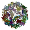
| Symmetry | Point symmetry: I (icosahedral) |
| 3D reconstruction | Resolution: 3.3 Å / Resolution method: FSC 0.143 CUT-OFF / Num. of particles: 4442 / Symmetry type: POINT |
