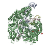
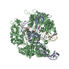
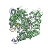
[English] 日本語
Yorodumi
- PDB-5z0b: Crystal structure of plasma-derived human serum albumin -

+
Open data


ID or keywords:

Loading...

-
Basic information

Entry
Database: PDB / ID: 5z0b
TitleCrystal structure of plasma-derived human serum albumin
ComponentsSerum albumin
KeywordsTRANSPORT PROTEIN / Human serum albumin
Function / homology
Function and homology information


cellular response to calcium ion starvation / exogenous protein binding / Ciprofloxacin ADME / HDL remodeling / enterobactin binding / Heme biosynthesis / negative regulation of mitochondrial depolarization / Prednisone ADME / Heme degradation / antioxidant activity ...cellular response to calcium ion starvation / exogenous protein binding / Ciprofloxacin ADME / HDL remodeling / enterobactin binding / Heme biosynthesis / negative regulation of mitochondrial depolarization / Prednisone ADME / Heme degradation / antioxidant activity / Aspirin ADME / toxic substance binding / Scavenging of heme from plasma / Recycling of bile acids and salts / cellular response to starvation / platelet alpha granule lumen / fatty acid binding / Post-translational protein phosphorylation / Cytoprotection by HMOX1 / Regulation of Insulin-like Growth Factor (IGF) transport and uptake by Insulin-like Growth Factor Binding Proteins (IGFBPs) / pyridoxal phosphate binding / Platelet degranulation / protein-folding chaperone binding / blood microparticle / copper ion binding / endoplasmic reticulum lumen / Golgi apparatus / endoplasmic reticulum / protein-containing complex / DNA binding / extracellular space / extracellular exosome / extracellular region / identical protein binding / nucleus / cytoplasm
Similarity search - Function
Serum Albumin; Chain A, Domain 1 - #10 / Serum albumin/Alpha-fetoprotein/Afamin / ALB/AFP/VDB / Serum albumin, N-terminal / Serum albumin, conserved site / Serum albumin-like / Serum albumin family / Albumin domain signature. / Albumin domain profile. / serum albumin ...Serum Albumin; Chain A, Domain 1 - #10 / Serum albumin/Alpha-fetoprotein/Afamin / ALB/AFP/VDB / Serum albumin, N-terminal / Serum albumin, conserved site / Serum albumin-like / Serum albumin family / Albumin domain signature. / Albumin domain profile. / serum albumin / Serum Albumin; Chain A, Domain 1 / Orthogonal Bundle / Mainly Alpha
Similarity search - Domain/homology
LINOLEIC ACID / OCTANOIC ACID (CAPRYLIC ACID) / DI(HYDROXYETHYL)ETHER / TRIETHYLENE GLYCOL / PALMITIC ACID / TRYPTOPHAN / Albumin
Similarity search - Component
Biological speciesHomo sapiens (human)
MethodX-RAY DIFFRACTION / SYNCHROTRON / MOLECULAR REPLACEMENT / Resolution: 2.17 Å
AuthorsPark, J. / Kim, M.-S. / Shin, D.H.
Funding support Korea, Republic Of, 1items
OrganizationGrant numberCountry
National Research Foundation (NRF, Korea)2015R1D1A1A01058942 Korea, Republic Of
CitationJournal: To Be Published
Title: The crystal structure of plasma-derived human serum albumin
Authors: Park, J. / Kim, M.S. / Shin, D.H.
History
DepositionDec 19, 2017Deposition site: PDBJ / Processing site: PDBJ
Revision 1.0Dec 19, 2018Provider: repository / Type: Initial release
Revision 2.0Jun 24, 2020Group: Advisory / Atomic model ...Advisory / Atomic model / Author supporting evidence / Data collection / Derived calculations / Non-polymer description / Other / Refinement description / Structure summary
Category: atom_site / atom_site_anisotrop ...atom_site / atom_site_anisotrop / cell / chem_comp / computing / diffrn / entity / pdbx_audit_support / pdbx_entity_instance_feature / pdbx_entity_nonpoly / pdbx_entry_details / pdbx_nonpoly_scheme / pdbx_refine_tls / pdbx_refine_tls_group / pdbx_struct_assembly_gen / pdbx_struct_assembly_prop / pdbx_validate_close_contact / pdbx_validate_symm_contact / pdbx_validate_torsion / refine / refine_hist / refine_ls_restr / refine_ls_shell / reflns / software / struct_asym / struct_conf / struct_conn / struct_mon_prot_cis / struct_site / struct_site_gen / symmetry
Item: _cell.angle_beta / _cell.volume ..._cell.angle_beta / _cell.volume / _chem_comp.formula / _chem_comp.formula_weight / _chem_comp.id / _chem_comp.mon_nstd_flag / _chem_comp.name / _chem_comp.pdbx_synonyms / _chem_comp.type / _diffrn.pdbx_serial_crystal_experiment / _pdbx_audit_support.funding_organization / _pdbx_struct_assembly_gen.asym_id_list / _pdbx_struct_assembly_prop.value / _pdbx_validate_symm_contact.auth_asym_id_1 / _pdbx_validate_symm_contact.auth_asym_id_2 / _pdbx_validate_symm_contact.auth_atom_id_1 / _pdbx_validate_symm_contact.auth_atom_id_2 / _pdbx_validate_symm_contact.auth_comp_id_1 / _pdbx_validate_symm_contact.auth_comp_id_2 / _pdbx_validate_symm_contact.auth_seq_id_1 / _pdbx_validate_symm_contact.auth_seq_id_2 / _pdbx_validate_symm_contact.dist / _pdbx_validate_symm_contact.site_symmetry_2 / _refine.B_iso_mean / _refine.ls_R_factor_R_free / _refine.ls_R_factor_R_work / _refine.ls_R_factor_obs / _refine.ls_d_res_high / _refine.ls_number_reflns_R_work / _refine.ls_number_reflns_all / _refine.overall_SU_ML / _refine.pdbx_overall_phase_error / _refine.pdbx_stereochemistry_target_values / _refine.solvent_model_details / _refine_hist.d_res_high / _refine_hist.number_atoms_solvent / _refine_hist.number_atoms_total / _refine_hist.pdbx_number_atoms_ligand / _refine_ls_restr.dev_ideal / _refine_ls_restr.number / _refine_ls_restr.type / _refine_ls_shell.R_factor_R_free / _refine_ls_shell.R_factor_R_work / _refine_ls_shell.d_res_high / _refine_ls_shell.d_res_low / _refine_ls_shell.number_reflns_R_free / _refine_ls_shell.number_reflns_R_work / _refine_ls_shell.percent_reflns_obs / _reflns.B_iso_Wilson_estimate / _software.version / _struct_mon_prot_cis.pdbx_omega_angle / _symmetry.space_group_name_Hall
Description: Ligand geometry
Details: We have replaced several fatty acid ligands with longer fatty acids, re-modeled the ligand geometry, and refined the overall protein structure.
Provider: author / Type: Coordinate replacement
Revision 2.1Nov 22, 2023Group: Data collection / Database references / Refinement description
Category: chem_comp_atom / chem_comp_bond ...chem_comp_atom / chem_comp_bond / database_2 / pdbx_initial_refinement_model
Item: _database_2.pdbx_DOI / _database_2.pdbx_database_accession

-
Structure visualization

Structure viewerMolecule:
MolmilJmol/JSmol

Downloads & links

-
Assembly

Deposited unit
A: Serum albumin
B: Serum albumin
C: Serum albumin
hetero molecules


Theoretical massNumber of molelcules
Total (without water)207,97760
Polymers199,7623
Non-polymers8,21657
Water8,305461
1


  • Idetical with deposited unit
  • defined by author
TypeNameSymmetry operationNumber
identity operation1_555x,y,z1
Buried area21180 Å2
ΔGint-160 kcal/mol
Surface area76450 Å2
Unit cell
Length a, b, c (Å)203.576, 113.466, 86.809
Angle α, β, γ (deg.)90.000, 104.878, 90.000
Int Tables number5
Space group name H-MC121
Space group name HallC2y

-
Components

-
Protein , 1 types, 3 molecules ABC

#1: Protein Serum albumin /


Mass: 66587.219 Da / Num. of mol.: 3 / Source method: isolated from a natural source / Source: (natural) Homo sapiens (human) / References: UniProt: P02768

-
Non-polymers , 10 types, 518 molecules

#2: Chemical ChemComp-EIC / LINOLEIC ACID / 9,12-LINOLEIC ACID / Linoleic acid


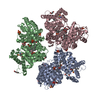
Mass: 280.445 Da / Num. of mol.: 3 / Source method: obtained synthetically / Formula: C18H32O2 / Feature type: SUBJECT OF INVESTIGATION
#3: Chemical
ChemComp-OCA / OCTANOIC ACID (CAPRYLIC ACID) / Caprylic acid


Mass: 144.211 Da / Num. of mol.: 5 / Source method: obtained synthetically / Formula: C8H16O2 / Feature type: SUBJECT OF INVESTIGATION
#4: Chemical
ChemComp-PLM / PALMITIC ACID / Palmitic acid


Mass: 256.424 Da / Num. of mol.: 4 / Source method: obtained synthetically / Formula: C16H32O2 / Feature type: SUBJECT OF INVESTIGATION
#5: Chemical ChemComp-TRP / TRYPTOPHAN / Tryptophan


Type: L-peptide linking / Mass: 204.225 Da / Num. of mol.: 3 / Source method: obtained synthetically / Formula: C11H12N2O2 / Feature type: SUBJECT OF INVESTIGATION
#6: Chemical
ChemComp-SO4 / SULFATE ION / Sulfate


Mass: 96.063 Da / Num. of mol.: 19 / Source method: isolated from a natural source / Formula: SO4
#7: Chemical
ChemComp-PEG / DI(HYDROXYETHYL)ETHER / Diethylene glycol


Mass: 106.120 Da / Num. of mol.: 13 / Source method: obtained synthetically / Formula: C4H10O3
#8: Chemical
ChemComp-PGE / TRIETHYLENE GLYCOL / Polyethylene glycol


Mass: 150.173 Da / Num. of mol.: 4 / Source method: obtained synthetically / Formula: C6H14O4
#9: Chemical
ChemComp-PG4 / TETRAETHYLENE GLYCOL / Polyethylene glycol


Mass: 194.226 Da / Num. of mol.: 5 / Source method: obtained synthetically / Formula: C8H18O5 / Comment: precipitant*YM
#10: Chemical ChemComp-1PE / PENTAETHYLENE GLYCOL / PEG400 / Polyethylene glycol


Mass: 238.278 Da / Num. of mol.: 1 / Source method: obtained synthetically / Formula: C10H22O6 / Comment: precipitant*YM
#11: Water ChemComp-HOH / water / Water


Mass: 18.015 Da / Num. of mol.: 461 / Source method: isolated from a natural source / Formula: H2O

-
Details

Has ligand of interestY

-
Experimental details

-
Experiment

ExperimentMethod: X-RAY DIFFRACTION / Number of used crystals: 1

-
Sample preparation

CrystalDensity Matthews: 2.46 Å3/Da / Density % sol: 49.99 %
Description: THE ENTRY CONTAINS FRIEDEL PAIRS IN F_PLUS/MINUS COLUMNS.
Crystal growTemperature: 296 K / Method: vapor diffusion, hanging drop / pH: 6.5 / Details: PEG600, Ammonium sulfate, MES

-
Data collection

DiffractionMean temperature: 100 K / Serial crystal experiment: N
Diffraction sourceSource: SYNCHROTRON / Site: Australian Synchrotron / Beamline: MX1 / Wavelength: 0.9536 Å
DetectorType: ADSC QUANTUM 315r / Detector: CCD / Date: Jul 18, 2011
RadiationProtocol: SINGLE WAVELENGTH / Monochromatic (M) / Laue (L): M / Scattering type: x-ray
Radiation wavelengthWavelength: 0.9536 Å / Relative weight: 1
ReflectionResolution: 2.17→19.78 Å / Num. obs: 96299 / % possible obs: 96.2 % / Redundancy: 7.1 % / Biso Wilson estimate: 35.86 Å2 / Rsym value: 0.053 / Net I/σ(I): 10.8
Reflection shellResolution: 2.17→2.29 Å / Mean I/σ(I) obs: 2.7 / Num. unique obs: 11416 / Rsym value: 0.29 / % possible all: 78.3

-
Processing

Software
NameVersionClassification
PHENIX1.16_3549refinement
XDSdata reduction
SCALAdata scaling
PHENIXphasing
RefinementMethod to determine structure: MOLECULAR REPLACEMENT
Starting model: 1AO6
Resolution: 2.17→19.78 Å / SU ML: 0.258 / Cross valid method: FREE R-VALUE / σ(F): 1.38 / Phase error: 24.3239
Stereochemistry target values: GeoStd + Monomer Library + CDL v1.2
Details: SF FILE CONTAINS FRIEDEL PAIRS UNDER I/F_MINUS AND I/F_PLUS COLUMNS.
RfactorNum. reflection% reflection
Rfree0.2323 4822 5.01 %
Rwork0.1828 91467 -
obs0.1853 96289 96.19 %
Solvent computationShrinkage radii: 0.9 Å / VDW probe radii: 1.11 Å / Solvent model: FLAT BULK SOLVENT MODEL
Displacement parametersBiso mean: 47.61 Å2
Refinement stepCycle: LAST / Resolution: 2.17→19.78 Å
ProteinNucleic acidLigandSolventTotal
Num. atoms13495 0 534 461 14490
Refine LS restraints
Refine-IDTypeDev idealNumber
X-RAY DIFFRACTIONf_bond_d0.007714268
X-RAY DIFFRACTIONf_angle_d0.914319109
X-RAY DIFFRACTIONf_chiral_restr0.04432067
X-RAY DIFFRACTIONf_plane_restr0.0072440
X-RAY DIFFRACTIONf_dihedral_angle_d17.4175644
LS refinement shell
Resolution (Å)Rfactor RfreeNum. reflection RfreeRfactor RworkNum. reflection RworkRefine-ID% reflection obs (%)
2.17-2.20.2775960.25251954X-RAY DIFFRACTION61.36
2.2-2.220.30521200.22582386X-RAY DIFFRACTION75.26
2.22-2.250.25741170.22132540X-RAY DIFFRACTION80.64
2.25-2.280.27291580.21672785X-RAY DIFFRACTION88.46
2.28-2.310.28471710.21093060X-RAY DIFFRACTION97.85
2.31-2.340.25041740.20383147X-RAY DIFFRACTION98.93
2.34-2.370.23361550.19873081X-RAY DIFFRACTION98.69
2.37-2.410.23651600.19493155X-RAY DIFFRACTION98.98
2.41-2.450.27021440.20123119X-RAY DIFFRACTION98.97
2.45-2.490.25471460.19823176X-RAY DIFFRACTION98.96
2.49-2.530.25781720.19993129X-RAY DIFFRACTION99.01
2.53-2.580.26671440.20013141X-RAY DIFFRACTION99.07
2.58-2.630.2721790.19613126X-RAY DIFFRACTION99.07
2.63-2.680.25111850.19653099X-RAY DIFFRACTION99.21
2.68-2.740.23121370.193154X-RAY DIFFRACTION99.1
2.74-2.80.24611740.20133174X-RAY DIFFRACTION99.23
2.8-2.870.25161770.19753089X-RAY DIFFRACTION99.15
2.87-2.950.24631750.20473123X-RAY DIFFRACTION99.34
2.95-3.030.2591460.20213165X-RAY DIFFRACTION99.43
3.03-3.130.27071550.20363175X-RAY DIFFRACTION99.34
3.13-3.240.2571670.19553172X-RAY DIFFRACTION99.38
3.24-3.370.25251460.19243153X-RAY DIFFRACTION99.49
3.37-3.530.22931700.17723149X-RAY DIFFRACTION99.55
3.53-3.710.21911990.16973116X-RAY DIFFRACTION99.49
3.71-3.940.20341940.16693156X-RAY DIFFRACTION99.7
3.94-4.240.20331700.15333163X-RAY DIFFRACTION99.55
4.24-4.660.20621580.15133193X-RAY DIFFRACTION99.73
4.66-5.330.22541870.1643170X-RAY DIFFRACTION99.76
5.33-6.670.22651700.19583205X-RAY DIFFRACTION99.94
6.67-19.780.20441760.16983212X-RAY DIFFRACTION99.01
Refinement TLS params.Method: refined / Origin x: 39.4380300071 Å / Origin y: -8.39593435405 Å / Origin z: -18.2087829151 Å
111213212223313233
T0.358170978783 Å2-0.00734545067059 Å20.00443158604637 Å2-0.356259378784 Å2-0.025069542985 Å2--0.304270145083 Å2
L0.183653343441 °20.0579389621211 °20.0173917792905 °2-0.289964508204 °2-0.105662211305 °2--0.0334615613146 °2
S-0.0284112655503 Å °0.0643034805569 Å °0.0113985798518 Å °-0.0854270752174 Å °0.000789223832073 Å °-0.0085150038951 Å °0.012200153297 Å °-0.0217686084264 Å °1.87666820287E-13 Å °
Refinement TLS groupSelection details: all

+
About Yorodumi

-
News

-
Feb 9, 2022. New format data for meta-information of EMDB entries

New format data for meta-information of EMDB entries

  • Version 3 of the EMDB header file is now the official format.
  • The previous official version 1.9 will be removed from the archive.

Related info.:EMDB header

External links:wwPDB to switch to version 3 of the EMDB data model

-
Aug 12, 2020. Covid-19 info

Covid-19 info

URL: https://pdbj.org/emnavi/covid19.php

New page: Covid-19 featured information page in EM Navigator.

Related info.:Covid-19 info / Mar 5, 2020. Novel coronavirus structure data

+
Mar 5, 2020. Novel coronavirus structure data

Novel coronavirus structure data

Related info.:Yorodumi Speices / Aug 12, 2020. Covid-19 info

External links:COVID-19 featured content - PDBj / Molecule of the Month (242):Coronavirus Proteases

+
Jan 31, 2019. EMDB accession codes are about to change! (news from PDBe EMDB page)

EMDB accession codes are about to change! (news from PDBe EMDB page)

  • The allocation of 4 digits for EMDB accession codes will soon come to an end. Whilst these codes will remain in use, new EMDB accession codes will include an additional digit and will expand incrementally as the available range of codes is exhausted. The current 4-digit format prefixed with “EMD-” (i.e. EMD-XXXX) will advance to a 5-digit format (i.e. EMD-XXXXX), and so on. It is currently estimated that the 4-digit codes will be depleted around Spring 2019, at which point the 5-digit format will come into force.
  • The EM Navigator/Yorodumi systems omit the EMD- prefix.

Related info.:Q: What is EMD? / ID/Accession-code notation in Yorodumi/EM Navigator

External links:EMDB Accession Codes are Changing Soon! / Contact to PDBj

+
Jul 12, 2017. Major update of PDB

Major update of PDB

  • wwPDB released updated PDB data conforming to the new PDBx/mmCIF dictionary.
  • This is a major update changing the version number from 4 to 5, and with Remediation, in which all the entries are updated.
  • In this update, many items about electron microscopy experimental information are reorganized (e.g. em_software).
  • Now, EM Navigator and Yorodumi are based on the updated data.

External links:wwPDB Remediation / Enriched Model Files Conforming to OneDep Data Standards Now Available in the PDB FTP Archive

-
Yorodumi

Thousand views of thousand structures

  • Yorodumi is a browser for structure data from EMDB, PDB, SASBDB, etc.
  • This page is also the successor to EM Navigator detail page, and also detail information page/front-end page for Omokage search.
  • The word "yorodu" (or yorozu) is an old Japanese word meaning "ten thousand". "mi" (miru) is to see.

Related info.:EMDB / PDB / SASBDB / Comparison of 3 databanks / Yorodumi Search / Aug 31, 2016. New EM Navigator & Yorodumi / Yorodumi Papers / Jmol/JSmol / Function and homology information / Changes in new EM Navigator and Yorodumi

Read more