 Movie
Movie Controller

[English] 日本語
 Yorodumi
- PDB-5yzn: Crystal structure of S9 peptidase (active form) from Deinococcus ... -
+ Open data

- Basic information
| Entry | Database: PDB / ID: 5yzn | ||||||
|---|---|---|---|---|---|---|---|
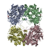
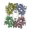
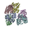
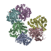
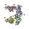
| Title | Crystal structure of S9 peptidase (active form) from Deinococcus radiodurans R1 | ||||||
 Components Components | Acyl-peptide hydrolase, putative | ||||||
 Keywords Keywords | HYDROLASE / Serine peptidase / Merops S9 / POP family | ||||||
| Function / homology |  Function and homology information Function and homology information | ||||||
| Biological species |  Deinococcus radiodurans (radioresistant) Deinococcus radiodurans (radioresistant) | ||||||
| Method |  X-RAY DIFFRACTION / SYNCHROTRON / MOLECULAR REPLACEMENT / Resolution: 2.3 Å X-RAY DIFFRACTION / SYNCHROTRON / MOLECULAR REPLACEMENT / Resolution: 2.3 Å | ||||||
 Authors Authors | Yadav, P. / Jamdar, S.N. / Kumar, A. / Ghosh, B. / Makde, R.D. | ||||||
 Citation Citation | Journal: J.Biol.Chem. / Year: 2019 Title: Carboxypeptidase in prolyl oligopeptidase family: Unique enzyme activation and substrate-screening mechanisms. Authors: Yadav, P. / Goyal, V.D. / Gaur, N.K. / Kumar, A. / Gokhale, S.M. / Jamdar, S.N. / Makde, R.D. | ||||||
| History |
|
- Structure visualization
| Structure viewer | Molecule: MolmilJmol/JSmol |
|---|
- Downloads & links
-Download
| PDBx/mmCIF format | 5yzn.cif.gz | 997.2 KB | Display | PDBx/mmCIF format |
|---|---|---|---|---|
| PDB format | pdb5yzn.ent.gz | 834.5 KB | Display | PDB format |
| PDBx/mmJSON format | 5yzn.json.gz | Tree view | PDBx/mmJSON format | |
| Others |  Other downloads Other downloads |
-Validation report
| Summary document | 5yzn_validation.pdf.gz | 459.4 KB | Display | wwPDB validaton report |
|---|---|---|---|---|
| Full document | 5yzn_full_validation.pdf.gz | 473.8 KB | Display | |
| Data in XML | 5yzn_validation.xml.gz | 97.1 KB | Display | |
| Data in CIF | 5yzn_validation.cif.gz | 138.8 KB | Display | |
| Arichive directory | https://data.pdbj.org/pub/pdb/validation_reports/yz/5yznftp://data.pdbj.org/pub/pdb/validation_reports/yz/5yzn | HTTPS FTP |
-Related structure data
| Related structure data | 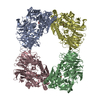 5yzmSC  5yzoC 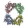 6igpC 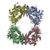 6igqC  6igrC  6ikgC S: Starting model for refinement C: citing same article ( |
|---|---|
| Similar structure data |


-Links
 PDBj
PDBj


- Assembly
| Deposited unit | 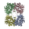
| ||||||||
|---|---|---|---|---|---|---|---|---|---|
| 1 |
| ||||||||
| Unit cell |
|
-Components
| #1: Protein | Mass: 72728.797 Da / Num. of mol.: 4 Source method: isolated from a genetically manipulated source Source: (gene. exp.) Deinococcus radiodurans (strain ATCC 13939 / DSM 20539 / JCM 16871 / LMG 4051 / NBRC 15346 / NCIMB 9279 / R1 / VKM B-1422) (radioresistant)Strain: ATCC 13939 / DSM 20539 / JCM 16871 / LMG 4051 / NBRC 15346 / NCIMB 9279 / R1 / VKM B-1422 Gene: DR_0165 / Plasmid: pST50Tr / Details (production host): pET based plasmid / Production host: #2: Water | ChemComp-HOH / |  Mass: 18.015 Da / Num. of mol.: 1331 / Source method: isolated from a natural source / Formula: H2O Mass: 18.015 Da / Num. of mol.: 1331 / Source method: isolated from a natural source / Formula: H2O |
|---|
-Experimental details
-Experiment
| Experiment | Method: X-RAY DIFFRACTION / Number of used crystals: 1 |
|---|
- Sample preparation
| Crystal | Density Matthews: 2.92 Å3/Da / Density % sol: 57.85 % / Description: parallelepiped, size 200-300 micron |
|---|---|
| Crystal grow | Temperature: 294 K / Method: microbatch / pH: 5.3 Details: 40mM potassium phosphate, 20% glycerol, 16% PEG 8000 PH range: 4.5-5.5 |
-Data collection
| Diffraction | Mean temperature: 100 K |
|---|---|
| Diffraction source | Source: SYNCHROTRON / Site: RRCAT INDUS-2  / Beamline: PX-BL21 / Wavelength: 0.97947 Å / Beamline: PX-BL21 / Wavelength: 0.97947 Å |
| Detector | Type: MARMOSAIC 225 mm CCD / Detector: CCD / Date: Nov 6, 2015 / Details: Mirrors |
| Radiation | Monochromator: Si 111 / Protocol: SINGLE WAVELENGTH / Monochromatic (M) / Laue (L): M / Scattering type: x-ray |
| Radiation wavelength | Wavelength: 0.97947 Å / Relative weight: 1 |
| Reflection | Resolution: 2.3→39.28 Å / Num. obs: 151191 / % possible obs: 100 % / Redundancy: 7.1 % / Biso Wilson estimate: 24.3 Å2 / CC1/2: 0.998 / Rmerge(I) obs: 0.124 / Rpim(I) all: 0.05 / Rrim(I) all: 0.133 / Net I/σ(I): 11.4 |
| Reflection shell | Resolution: 2.3→2.34 Å / Redundancy: 7.2 % / Rmerge(I) obs: 0.929 / Mean I/σ(I) obs: 2.1 / Num. unique obs: 7386 / CC1/2: 0.913 / Rpim(I) all: 0.373 / Rrim(I) all: 1.002 / % possible all: 99.9 |
- Processing
| Software |
| |||||||||||||||||||||||||||||||||||||||||||||||||||||||||||||||||||||||||||||||||||||||||||||||||||||||||||||||||||||||||||||||||||||||||||||||||||||||||||||||||||||||||||||||||||||||||||||||||||||||||||||||||||||||||
|---|---|---|---|---|---|---|---|---|---|---|---|---|---|---|---|---|---|---|---|---|---|---|---|---|---|---|---|---|---|---|---|---|---|---|---|---|---|---|---|---|---|---|---|---|---|---|---|---|---|---|---|---|---|---|---|---|---|---|---|---|---|---|---|---|---|---|---|---|---|---|---|---|---|---|---|---|---|---|---|---|---|---|---|---|---|---|---|---|---|---|---|---|---|---|---|---|---|---|---|---|---|---|---|---|---|---|---|---|---|---|---|---|---|---|---|---|---|---|---|---|---|---|---|---|---|---|---|---|---|---|---|---|---|---|---|---|---|---|---|---|---|---|---|---|---|---|---|---|---|---|---|---|---|---|---|---|---|---|---|---|---|---|---|---|---|---|---|---|---|---|---|---|---|---|---|---|---|---|---|---|---|---|---|---|---|---|---|---|---|---|---|---|---|---|---|---|---|---|---|---|---|---|---|---|---|---|---|---|---|---|---|---|---|---|---|---|---|---|
| Refinement | Method to determine structure: MOLECULAR REPLACEMENT Starting model: 5YZM Resolution: 2.3→39.279 Å / SU ML: 0.25 / Cross valid method: FREE R-VALUE / σ(F): 1.33 / Phase error: 31.68
| |||||||||||||||||||||||||||||||||||||||||||||||||||||||||||||||||||||||||||||||||||||||||||||||||||||||||||||||||||||||||||||||||||||||||||||||||||||||||||||||||||||||||||||||||||||||||||||||||||||||||||||||||||||||||
| Solvent computation | Shrinkage radii: 0.9 Å / VDW probe radii: 1.11 Å | |||||||||||||||||||||||||||||||||||||||||||||||||||||||||||||||||||||||||||||||||||||||||||||||||||||||||||||||||||||||||||||||||||||||||||||||||||||||||||||||||||||||||||||||||||||||||||||||||||||||||||||||||||||||||
| Refinement step | Cycle: LAST / Resolution: 2.3→39.279 Å
| |||||||||||||||||||||||||||||||||||||||||||||||||||||||||||||||||||||||||||||||||||||||||||||||||||||||||||||||||||||||||||||||||||||||||||||||||||||||||||||||||||||||||||||||||||||||||||||||||||||||||||||||||||||||||
| Refine LS restraints |
| |||||||||||||||||||||||||||||||||||||||||||||||||||||||||||||||||||||||||||||||||||||||||||||||||||||||||||||||||||||||||||||||||||||||||||||||||||||||||||||||||||||||||||||||||||||||||||||||||||||||||||||||||||||||||
| LS refinement shell |
| |||||||||||||||||||||||||||||||||||||||||||||||||||||||||||||||||||||||||||||||||||||||||||||||||||||||||||||||||||||||||||||||||||||||||||||||||||||||||||||||||||||||||||||||||||||||||||||||||||||||||||||||||||||||||
| Refinement TLS params. | Method: refined / Origin x: 132.5933 Å / Origin y: 173.8118 Å / Origin z: 47.5903 Å
| |||||||||||||||||||||||||||||||||||||||||||||||||||||||||||||||||||||||||||||||||||||||||||||||||||||||||||||||||||||||||||||||||||||||||||||||||||||||||||||||||||||||||||||||||||||||||||||||||||||||||||||||||||||||||
| Refinement TLS group | Selection details: all |
