 Movie
Movie Controller

[English] 日本語
 Yorodumi
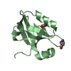
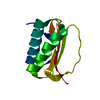
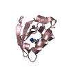
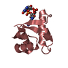
- PDB-5ytt: Crystal structure of YB1 cold-shock domain in complex with UCAUGU -
+ Open data

- Basic information
| Entry | Database: PDB / ID: 5ytt | |||||||||
|---|---|---|---|---|---|---|---|---|---|---|
| Title | Crystal structure of YB1 cold-shock domain in complex with UCAUGU | |||||||||
 Components Components |
| |||||||||
 Keywords Keywords | RNA BINDING PROTEIN/RNA / YB1 / cold-shock domain / CAUG / RNA BINDING PROTEIN / RNA BINDING PROTEIN-RNA complex | |||||||||
| Function / homology |  Function and homology information Function and homology informationtRNA transport / CRD-mediated mRNA stability complex / C5-methylcytidine-containing RNA reader activity / miRNA transport / negative regulation of striated muscle cell differentiation / negative regulation of nuclear-transcribed mRNA catabolic process, deadenylation-dependent decay / Noncanonical activation of NOTCH3 / CRD-mediated mRNA stabilization / protein localization to cytoplasmic stress granule / RNA transport ...tRNA transport / CRD-mediated mRNA stability complex / C5-methylcytidine-containing RNA reader activity / miRNA transport / negative regulation of striated muscle cell differentiation / negative regulation of nuclear-transcribed mRNA catabolic process, deadenylation-dependent decay / Noncanonical activation of NOTCH3 / CRD-mediated mRNA stabilization / protein localization to cytoplasmic stress granule / RNA transport / histone pre-mRNA 3'end processing complex / embryonic morphogenesis / U12-type spliceosomal complex / positive regulation of cytoplasmic translation / cellular response to interleukin-7 / miRNA binding / mRNA stabilization / mRNA Splicing - Minor Pathway / Processing of Capped Intron-Containing Pre-mRNA / positive regulation of cell division / negative regulation of cellular senescence / epidermis development / mRNA Splicing - Major Pathway / RNA splicing / P-body / SMAD2/SMAD3:SMAD4 heterotrimer regulates transcription / Interferon gamma signaling / mRNA processing / cytoplasmic stress granule / sequence-specific double-stranded DNA binding / single-stranded DNA binding / regulation of gene expression / double-stranded DNA binding / GTPase binding / in utero embryonic development / nucleic acid binding / negative regulation of translation / ribonucleoprotein complex / mRNA binding / chromatin binding / regulation of DNA-templated transcription / synapse / negative regulation of transcription by RNA polymerase II / endoplasmic reticulum / positive regulation of transcription by RNA polymerase II / DNA binding / RNA binding / extracellular exosome / extracellular region / nucleoplasm / nucleus / plasma membrane / cytoplasm / cytosol Similarity search - Function | |||||||||
| Biological species |  Homo sapiens (human) Homo sapiens (human)synthetic construct (others) | |||||||||
| Method |  X-RAY DIFFRACTION / SYNCHROTRON / MOLECULAR REPLACEMENT / Resolution: 1.6 Å X-RAY DIFFRACTION / SYNCHROTRON / MOLECULAR REPLACEMENT / Resolution: 1.6 Å | |||||||||
 Authors Authors | Yang, X. / Huang, Y. | |||||||||
| Funding support |  China, 2items China, 2items
| |||||||||
 Citation Citation | Journal: J.Biol.Chem. / Year: 2019 Title: Crystal structure of a Y-box binding protein 1 (YB-1)-RNA complex reveals key features and residues interacting with RNA. Authors: Yang, X.J. / Zhu, H. / Mu, S.R. / Wei, W.J. / Yuan, X. / Wang, M. / Liu, Y. / Hui, J. / Huang, Y. | |||||||||
| History |
|
- Structure visualization
| Structure viewer | Molecule: MolmilJmol/JSmol |
|---|
- Downloads & links
-Download
| PDBx/mmCIF format | 5ytt.cif.gz | 57.4 KB | Display | PDBx/mmCIF format |
|---|---|---|---|---|
| PDB format | pdb5ytt.ent.gz | 38.3 KB | Display | PDB format |
| PDBx/mmJSON format | 5ytt.json.gz | Tree view | PDBx/mmJSON format | |
| Others |  Other downloads Other downloads |
-Validation report
| Arichive directory | https://data.pdbj.org/pub/pdb/validation_reports/yt/5yttftp://data.pdbj.org/pub/pdb/validation_reports/yt/5ytt | HTTPS FTP |
|---|
-Related structure data
| Related structure data | 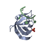 5ytsC 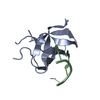 5ytvC  5ytxC 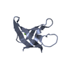 3pf5S C: citing same article ( S: Starting model for refinement |
|---|---|
| Similar structure data |


-Links
 PDBj
PDBj


- Assembly
| Deposited unit | 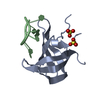
| ||||||||
|---|---|---|---|---|---|---|---|---|---|
| 1 |
| ||||||||
| Unit cell |
|
-Components
| #1: Protein | Mass: 8997.026 Da / Num. of mol.: 1 Source method: isolated from a genetically manipulated source Source: (gene. exp.) Homo sapiens (human) / Gene: YBX1, NSEP1, YB1 / Production host:  | ||
|---|---|---|---|
| #2: RNA chain | Mass: 2159.299 Da / Num. of mol.: 1 / Source method: obtained synthetically / Source: (synth.) synthetic construct (others) | ||
| #3: Chemical |   Mass: 96.063 Da / Num. of mol.: 2 / Source method: obtained synthetically / Formula: SO4 Mass: 96.063 Da / Num. of mol.: 2 / Source method: obtained synthetically / Formula: SO4#4: Water | ChemComp-HOH / |  Mass: 18.015 Da / Num. of mol.: 62 / Source method: isolated from a natural source / Formula: H2O Mass: 18.015 Da / Num. of mol.: 62 / Source method: isolated from a natural source / Formula: H2O |
-Experimental details
-Experiment
| Experiment | Method: X-RAY DIFFRACTION / Number of used crystals: 1 |
|---|
- Sample preparation
| Crystal | Density Matthews: 2.1 Å3/Da / Density % sol: 41.56 % |
|---|---|
| Crystal grow | Temperature: 290 K / Method: vapor diffusion, hanging drop / pH: 4.5 Details: 30 % (v/v) 2-methyl-2,4-pentanediol, 0.1 M sodium acetate, pH 4.5, 25 % (w/v) PEG 1500 |
-Data collection
| Diffraction | Mean temperature: 100 K |
|---|---|
| Diffraction source | Source: SYNCHROTRON / Site: SSRF / Beamline: BL17U1 / Wavelength: 0.97852 Å |
| Detector | Type: ADSC QUANTUM 315 / Detector: CCD / Date: Dec 12, 2014 |
| Radiation | Protocol: SINGLE WAVELENGTH / Monochromatic (M) / Laue (L): M / Scattering type: x-ray |
| Radiation wavelength | Wavelength: 0.97852 Å / Relative weight: 1 |
| Reflection | Resolution: 1.6→50 Å / Num. obs: 11696 / % possible obs: 96.4 % / Redundancy: 9.2 % / Net I/σ(I): 26.6 |
| Reflection shell | Resolution: 1.6→1.66 Å |
- Processing
| Software |
| |||||||||||||||||||||||||||||||||||||||||||||||||||||||||||||||
|---|---|---|---|---|---|---|---|---|---|---|---|---|---|---|---|---|---|---|---|---|---|---|---|---|---|---|---|---|---|---|---|---|---|---|---|---|---|---|---|---|---|---|---|---|---|---|---|---|---|---|---|---|---|---|---|---|---|---|---|---|---|---|---|---|
| Refinement | Method to determine structure: MOLECULAR REPLACEMENT Starting model: 3PF5 Resolution: 1.6→30 Å / SU ML: 0.17 / Cross valid method: FREE R-VALUE / σ(F): 1.36 / Phase error: 22.64
| |||||||||||||||||||||||||||||||||||||||||||||||||||||||||||||||
| Solvent computation | Shrinkage radii: 0.9 Å / VDW probe radii: 1.11 Å | |||||||||||||||||||||||||||||||||||||||||||||||||||||||||||||||
| Refinement step | Cycle: LAST / Resolution: 1.6→30 Å
| |||||||||||||||||||||||||||||||||||||||||||||||||||||||||||||||
| Refine LS restraints |
| |||||||||||||||||||||||||||||||||||||||||||||||||||||||||||||||
| LS refinement shell |
| |||||||||||||||||||||||||||||||||||||||||||||||||||||||||||||||
| Refinement TLS params. | Method: refined / Origin x: -6.2485 Å / Origin y: 23.1717 Å / Origin z: 36.1764 Å
| |||||||||||||||||||||||||||||||||||||||||||||||||||||||||||||||
| Refinement TLS group | Selection details: all |
