 Movie
Movie Controller

[English] 日本語
 Yorodumi
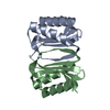
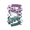
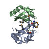
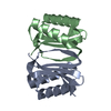
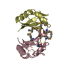
- PDB-3rjs: Crystal structure of Dynein Light Chain 8a (DLC8) from Toxoplasma... -
+ Open data

- Basic information
| Entry | Database: PDB / ID: 3rjs | ||||||
|---|---|---|---|---|---|---|---|
| Title | Crystal structure of Dynein Light Chain 8a (DLC8) from Toxoplasma gondii at 1.5 A resolution | ||||||
 Components Components | Dynein light chain motor protein | ||||||
 Keywords Keywords | MOTOR PROTEIN / Parasite / dynein light chain / LC8 / DLC8 / TgDLC8 / DYNEIN / LIGHT CHAIN / PIN / DLC1 / DYNLL1 / TRANSPORT PROTEIN | ||||||
| Function / homology |  Function and homology information Function and homology informationcytoplasmic dynein complex / dynein intermediate chain binding / microtubule-based process / microtubule Similarity search - Function | ||||||
| Biological species |  | ||||||
| Method |  X-RAY DIFFRACTION / SYNCHROTRON / MOLECULAR REPLACEMENT / Resolution: 1.5 Å X-RAY DIFFRACTION / SYNCHROTRON / MOLECULAR REPLACEMENT / Resolution: 1.5 Å | ||||||
 Authors Authors | Qureshi, B. / Hoehne, W. / Scheerer, P. | ||||||
 Citation Citation | Journal: Faseb J. / Year: 2013 Title: Dynein light chain 8a of Toxoplasma gondii, a unique conoid-localized beta-strand-swapped homodimer, is required for an efficient parasite growth. Authors: Qureshi, B.M. / Hofmann, N.E. / Arroyo-Olarte, R.D. / Nickl, B. / Hoehne, W. / Jungblut, P.R. / Lucius, R. / Scheerer, P. / Gupta, N. | ||||||
| History |
|
- Structure visualization
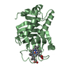
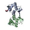
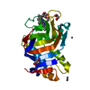
| Structure viewer | Molecule: MolmilJmol/JSmol |
|---|
- Downloads & links
-Download
| PDBx/mmCIF format | 3rjs.cif.gz | 51.9 KB | Display | PDBx/mmCIF format |
|---|---|---|---|---|
| PDB format | pdb3rjs.ent.gz | 37.6 KB | Display | PDB format |
| PDBx/mmJSON format | 3rjs.json.gz | Tree view | PDBx/mmJSON format | |
| Others |  Other downloads Other downloads |
-Validation report
| Arichive directory | https://data.pdbj.org/pub/pdb/validation_reports/rj/3rjsftp://data.pdbj.org/pub/pdb/validation_reports/rj/3rjs | HTTPS FTP |
|---|
-Related structure data
| Related structure data |  1cmiS S: Starting model for refinement |
|---|---|
| Similar structure data |


-Links
 PDBj
PDBj
- Assembly
| Deposited unit | 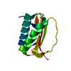
| ||||||||
|---|---|---|---|---|---|---|---|---|---|
| 1 | 
| ||||||||
| Unit cell |
| ||||||||
| Components on special symmetry positions |
|
-Components
| #1: Protein | Mass: 10317.735 Da / Num. of mol.: 1 Source method: isolated from a genetically manipulated source Source: (gene. exp.)  |
|---|---|
| #2: Water | ChemComp-HOH /  Mass: 18.015 Da / Num. of mol.: 113 / Source method: isolated from a natural source / Formula: H2O Mass: 18.015 Da / Num. of mol.: 113 / Source method: isolated from a natural source / Formula: H2O |
-Experimental details
-Experiment
| Experiment | Method: X-RAY DIFFRACTION / Number of used crystals: 1 |
|---|
- Sample preparation
| Crystal | Density Matthews: 2.23 Å3/Da / Density % sol: 44.87 % |
|---|---|
| Crystal grow | Temperature: 295 K / pH: 4.6 Details: 16% polyethylene glycol 4000, 0.2 M (NH4)2SO4, 0.1 M CH3COONa, pH 4.6, VAPOR DIFFUSION, HANGING DROP, temperature 295K |
-Data collection
| Diffraction | Mean temperature: 100 K |
|---|---|
| Diffraction source | Source: SYNCHROTRON / Site: BESSY  / Beamline: 14.2 / Wavelength: 0.9184 / Beamline: 14.2 / Wavelength: 0.9184 |
| Detector | Type: MAR CCD 165 mm / Detector: CCD / Date: May 23, 2007 / Details: MIRRORS |
| Radiation | Monochromator: SI-111 CRYSTAL - DOUBLE CRYSTAL MONOCHROMATOR Protocol: SINGLE WAVELENGTH / Monochromatic (M) / Laue (L): M / Scattering type: x-ray |
| Radiation wavelength | Wavelength: 0.9184 Å / Relative weight: 1 |
| Reflection | Resolution: 1.5→51.62 Å / Num. obs: 14931 / % possible obs: 99.7 % / Observed criterion σ(I): 0 / Redundancy: 6.1 % / Biso Wilson estimate: 23.8 Å2 / Rmerge(I) obs: 0.035 / Rsym value: 0.035 / Net I/σ(I): 46 |
| Reflection shell | Resolution: 1.5→1.53 Å / Redundancy: 6 % / Rmerge(I) obs: 0.252 / Mean I/σ(I) obs: 6.2 / Rsym value: 0.252 / % possible all: 100 |
- Processing
| Software |
| ||||||||||||||||||||||||||||||||||||||||||||||||||||||||||||||||||||||||||||||||||||||||||||||||||||
|---|---|---|---|---|---|---|---|---|---|---|---|---|---|---|---|---|---|---|---|---|---|---|---|---|---|---|---|---|---|---|---|---|---|---|---|---|---|---|---|---|---|---|---|---|---|---|---|---|---|---|---|---|---|---|---|---|---|---|---|---|---|---|---|---|---|---|---|---|---|---|---|---|---|---|---|---|---|---|---|---|---|---|---|---|---|---|---|---|---|---|---|---|---|---|---|---|---|---|---|---|---|
| Refinement | Method to determine structure: MOLECULAR REPLACEMENT Starting model: PDB ENTRY 1CMI Resolution: 1.5→18.756 Å / Cor.coef. Fo:Fc: 0.967 / Cor.coef. Fo:Fc free: 0.952 / SU ML: 0.13 / σ(F): 1.36 / Phase error: 22.88 / Stereochemistry target values: ML / Details: HYDROGENS HAVE BEEN ADDED IN THE RIDING POSITIONS
| ||||||||||||||||||||||||||||||||||||||||||||||||||||||||||||||||||||||||||||||||||||||||||||||||||||
| Solvent computation | Shrinkage radii: 0.9 Å / VDW probe radii: 1.11 Å / Solvent model: FLAT BULK SOLVENT MODEL | ||||||||||||||||||||||||||||||||||||||||||||||||||||||||||||||||||||||||||||||||||||||||||||||||||||
| Displacement parameters | Biso mean: 26.147 Å2
| ||||||||||||||||||||||||||||||||||||||||||||||||||||||||||||||||||||||||||||||||||||||||||||||||||||
| Refine analyze |
| ||||||||||||||||||||||||||||||||||||||||||||||||||||||||||||||||||||||||||||||||||||||||||||||||||||
| Refinement step | Cycle: LAST / Resolution: 1.5→18.756 Å
| ||||||||||||||||||||||||||||||||||||||||||||||||||||||||||||||||||||||||||||||||||||||||||||||||||||
| Refine LS restraints |
| ||||||||||||||||||||||||||||||||||||||||||||||||||||||||||||||||||||||||||||||||||||||||||||||||||||
| LS refinement shell |
| ||||||||||||||||||||||||||||||||||||||||||||||||||||||||||||||||||||||||||||||||||||||||||||||||||||
| Refinement TLS params. | Method: refined / Refine-ID: X-RAY DIFFRACTION
| ||||||||||||||||||||||||||||||||||||||||||||||||||||||||||||||||||||||||||||||||||||||||||||||||||||
| Refinement TLS group |
|
