 Movie
Movie Controller

[English] 日本語
 Yorodumi
- PDB-2ek1: Crystal structure of RNA-binding motif of human rna-binding protein 12 -
+ Open data

- Basic information
| Entry | Database: PDB / ID: 2ek1 | ||||||
|---|---|---|---|---|---|---|---|
| Title | Crystal structure of RNA-binding motif of human rna-binding protein 12 | ||||||
 Components Components | RNA-binding protein 12 | ||||||
 Keywords Keywords | RNA BINDING PROTEIN / RNA recognition motif / dimer / Structural Genomics / NPPSFA / National Project on Protein Structural and Functional Analyses / RIKEN Structural Genomics/Proteomics Initiative / RSGI | ||||||
| Function / homology |  Function and homology information Function and homology informationregulation of RNA splicing / ribonucleoprotein complex / RNA binding / nucleoplasm Similarity search - Function | ||||||
| Biological species |  Homo sapiens (human) Homo sapiens (human) | ||||||
| Method |  X-RAY DIFFRACTION / SYNCHROTRON / SAD / Resolution: 2 Å X-RAY DIFFRACTION / SYNCHROTRON / SAD / Resolution: 2 Å | ||||||
 Authors Authors | Ihsanawati / Bessho, Y. / Shirouzu, M. / Yokoyama, S. / RIKEN Structural Genomics/Proteomics Initiative (RSGI) | ||||||
 Citation Citation | Journal: To be Published Title: Crystal structure of RNA-binding motif of human rna-binding protein 12 Authors: Ihsanawati / Bessho, Y. / Shirouzu, M. / Yokoyama, S. | ||||||
| History |
|
- Structure visualization
| Structure viewer | Molecule: MolmilJmol/JSmol |
|---|
- Downloads & links
-Download
| PDBx/mmCIF format | 2ek1.cif.gz | 140.5 KB | Display | PDBx/mmCIF format |
|---|---|---|---|---|
| PDB format | pdb2ek1.ent.gz | 112.4 KB | Display | PDB format |
| PDBx/mmJSON format | 2ek1.json.gz | Tree view | PDBx/mmJSON format | |
| Others |  Other downloads Other downloads |
-Validation report
| Arichive directory | https://data.pdbj.org/pub/pdb/validation_reports/ek/2ek1ftp://data.pdbj.org/pub/pdb/validation_reports/ek/2ek1 | HTTPS FTP |
|---|
-Related structure data
| Similar structure data | |
|---|---|
| Other databases |


-Links
 PDBj
PDBj- Assembly
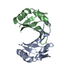
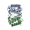
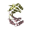
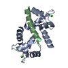
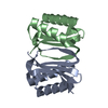
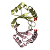
| Deposited unit | 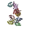
| ||||||||
|---|---|---|---|---|---|---|---|---|---|
| 1 | 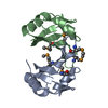
| ||||||||
| 2 | 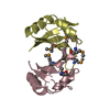
| ||||||||
| 3 | 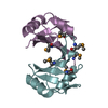
| ||||||||
| 4 | 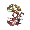
| ||||||||
| Unit cell |
|
-Components
| #1: Protein | Mass: 10067.821 Da / Num. of mol.: 8 / Fragment: residues 861-955 Source method: isolated from a genetically manipulated source Source: (gene. exp.) Homo sapiens (human) / Description: Cell Free system / Gene: RBM12, KIAA0765 / Plasmid: PX041122-21 / References: UniProt: Q9NTZ6#2: Water | ChemComp-HOH / |  Mass: 18.015 Da / Num. of mol.: 439 / Source method: isolated from a natural source / Formula: H2O Mass: 18.015 Da / Num. of mol.: 439 / Source method: isolated from a natural source / Formula: H2OHas protein modification | Y | |
|---|
-Experimental details
-Experiment
| Experiment | Method: X-RAY DIFFRACTION / Number of used crystals: 1 |
|---|
- Sample preparation
| Crystal | Density Matthews: 1.91 Å3/Da / Density % sol: 35.47 % |
|---|---|
| Crystal grow | Temperature: 293 K / Method: vapor diffusion, sitting drop / pH: 7.5 Details: 0.1M Hepes pH7.5, 25% (w/v) PEG 3000, VAPOR DIFFUSION, SITTING DROP, temperature 293K |
-Data collection
| Diffraction | Mean temperature: 100 K | ||||||||||||
|---|---|---|---|---|---|---|---|---|---|---|---|---|---|
| Diffraction source | Source: SYNCHROTRON / Site: Photon Factory  / Beamline: BL-5A / Wavelength: 0.97947, 0.97964, 0.964 / Beamline: BL-5A / Wavelength: 0.97947, 0.97964, 0.964 | ||||||||||||
| Detector | Type: ADSC QUANTUM 315 / Detector: CCD / Date: Dec 16, 2006 | ||||||||||||
| Radiation | Monochromator: Si / Protocol: MAD / Monochromatic (M) / Laue (L): M / Scattering type: x-ray | ||||||||||||
| Radiation wavelength |
| ||||||||||||
| Reflection | Resolution: 2→30 Å / Num. obs: 39769 / % possible obs: 97.1 % / Observed criterion σ(I): -3 / Redundancy: 3.6 % / Biso Wilson estimate: 19.7 Å2 / Rsym value: 0.102 | ||||||||||||
| Reflection shell | Resolution: 2→2.07 Å / Rsym value: 0.312 / % possible all: 89.8 |
- Processing
| Software |
| ||||||||||||||||||||
|---|---|---|---|---|---|---|---|---|---|---|---|---|---|---|---|---|---|---|---|---|---|
| Refinement | Method to determine structure: SAD / Resolution: 2→29.76 Å / Rfactor Rfree error: 0.006 / Data cutoff high absF: 1922580.3 / Data cutoff low absF: 0 / Isotropic thermal model: RESTRAINED / Cross valid method: THROUGHOUT / σ(F): 0 / Stereochemistry target values: Engh & Huber
| ||||||||||||||||||||
| Solvent computation | Solvent model: FLAT MODEL / Bsol: 42.3441 Å2 / ksol: 0.322088 e/Å3 | ||||||||||||||||||||
| Displacement parameters | Biso mean: 30.3 Å2
| ||||||||||||||||||||
| Refine analyze |
| ||||||||||||||||||||
| Refinement step | Cycle: LAST / Resolution: 2→29.76 Å
| ||||||||||||||||||||
| Refine LS restraints |
| ||||||||||||||||||||
| LS refinement shell | Resolution: 2→2.13 Å / Rfactor Rfree error: 0.018 / Total num. of bins used: 6
| ||||||||||||||||||||
| Xplor file |
|
