 Movie
Movie Controller

[English] 日本語
 Yorodumi
- PDB-5yk2: The complex structure of Rv3197-erythromycin from Mycobacterium t... -
+ Open data

- Basic information
| Entry | Database: PDB / ID: 5yk2 | ||||||||||||
|---|---|---|---|---|---|---|---|---|---|---|---|---|---|
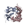
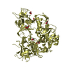
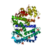
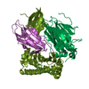
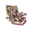
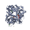
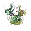
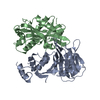
| Title | The complex structure of Rv3197-erythromycin from Mycobacterium tuberculosis | ||||||||||||
 Components Components | Probable conserved ATP-binding protein ABC transporter | ||||||||||||
 Keywords Keywords | TRANSPORT PROTEIN / Mycobacterium tuberculosis / macrolide antibiotic binding protein / non-canonical ABC protein | ||||||||||||
| Function / homology |  Function and homology information Function and homology information | ||||||||||||
| Biological species |   Mycobacterium tuberculosis (bacteria) Mycobacterium tuberculosis (bacteria) | ||||||||||||
| Method |  X-RAY DIFFRACTION / SYNCHROTRON / MOLECULAR REPLACEMENT / Resolution: 2.807 Å X-RAY DIFFRACTION / SYNCHROTRON / MOLECULAR REPLACEMENT / Resolution: 2.807 Å | ||||||||||||
 Authors Authors | Rao, Z.H. / Zhang, Q.Q. | ||||||||||||
| Funding support |  China, 3items China, 3items
| ||||||||||||
 Citation Citation | Journal: Protein Cell / Year: 2018 Title: Discovery of the first macrolide antibiotic binding protein in Mycobacterium tuberculosis: a new antibiotic resistance drug target. Authors: Zhang, Q. / Liu, H. / Liu, X. / Jiang, D. / Zhang, B. / Tian, H. / Yang, C. / Guddat, L.W. / Yang, H. / Mi, K. / Rao, Z. | ||||||||||||
| History |
|
- Structure visualization
| Structure viewer | Molecule: MolmilJmol/JSmol |
|---|
- Downloads & links
-Download
| PDBx/mmCIF format | 5yk2.cif.gz | 169.9 KB | Display | PDBx/mmCIF format |
|---|---|---|---|---|
| PDB format | pdb5yk2.ent.gz | 133.1 KB | Display | PDB format |
| PDBx/mmJSON format | 5yk2.json.gz | Tree view | PDBx/mmJSON format | |
| Others |  Other downloads Other downloads |
-Validation report
| Arichive directory | https://data.pdbj.org/pub/pdb/validation_reports/yk/5yk2ftp://data.pdbj.org/pub/pdb/validation_reports/yk/5yk2 | HTTPS FTP |
|---|
-Related structure data
| Related structure data | 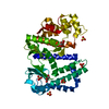 5yjzSC  5yk0C  5yk1C S: Starting model for refinement C: citing same article ( |
|---|---|
| Similar structure data |


-Links
 PDBj
PDBj

- Assembly
| Deposited unit | 
| ||||||||
|---|---|---|---|---|---|---|---|---|---|
| 1 |
| ||||||||
| Unit cell |
|
-Components
| #1: Protein | Mass: 49599.285 Da / Num. of mol.: 1 Source method: isolated from a genetically manipulated source Source: (gene. exp.) Mycobacterium tuberculosis (strain ATCC 25618 / H37Rv) (bacteria)Strain: ATCC 25618 / H37Rv / Gene: Rv3197 / Plasmid: pGEX-6P-1 / Production host: |
|---|---|
| #2: Chemical | ChemComp-ERY /   Mass: 733.927 Da / Num. of mol.: 1 / Source method: obtained synthetically / Formula: C37H67NO13 / Feature type: SUBJECT OF INVESTIGATION / Comment: antibiotic*YM Mass: 733.927 Da / Num. of mol.: 1 / Source method: obtained synthetically / Formula: C37H67NO13 / Feature type: SUBJECT OF INVESTIGATION / Comment: antibiotic*YM |
-Experimental details
-Experiment
| Experiment | Method: X-RAY DIFFRACTION / Number of used crystals: 1 |
|---|
- Sample preparation
| Crystal | Density Matthews: 3.47 Å3/Da / Density % sol: 64.5 % |
|---|---|
| Crystal grow | Temperature: 293 K / Method: vapor diffusion, sitting drop / pH: 6.5 Details: 1.8M ammonium sulfate, 0.1M Bis-Tris (pH 6.5), 2% (v/v) polyethylene glycol monomethyl ether 550 |
-Data collection
| Diffraction | Mean temperature: 100 K |
|---|---|
| Diffraction source | Source: SYNCHROTRON / Site: SSRF / Beamline: BL19U1 / Wavelength: 0.97855 Å |
| Detector | Type: DECTRIS PILATUS 6M / Detector: PIXEL / Date: Oct 9, 2014 |
| Radiation | Protocol: SINGLE WAVELENGTH / Monochromatic (M) / Laue (L): M / Scattering type: x-ray |
| Radiation wavelength | Wavelength: 0.97855 Å / Relative weight: 1 |
| Reflection | Resolution: 2.8→50 Å / Num. obs: 17350 / % possible obs: 99.4 % / Redundancy: 13.2 % / Rmerge(I) obs: 0.079 / Net I/σ(I): 26.5 |
| Reflection shell | Resolution: 2.8→2.9 Å / Redundancy: 13.4 % / Rmerge(I) obs: 0.723 / Mean I/σ(I) obs: 2.7 / Num. unique obs: 1686 / % possible all: 100 |
- Processing
| Software |
| |||||||||||||||||||||||||||||||||||||||||||||||||
|---|---|---|---|---|---|---|---|---|---|---|---|---|---|---|---|---|---|---|---|---|---|---|---|---|---|---|---|---|---|---|---|---|---|---|---|---|---|---|---|---|---|---|---|---|---|---|---|---|---|---|
| Refinement | Method to determine structure: MOLECULAR REPLACEMENT Starting model: 5YJZ Resolution: 2.807→34.712 Å / SU ML: 0.37 / Cross valid method: FREE R-VALUE / σ(F): 1.35 / Phase error: 27.1
| |||||||||||||||||||||||||||||||||||||||||||||||||
| Solvent computation | Shrinkage radii: 0.9 Å / VDW probe radii: 1.11 Å | |||||||||||||||||||||||||||||||||||||||||||||||||
| Refinement step | Cycle: LAST / Resolution: 2.807→34.712 Å
| |||||||||||||||||||||||||||||||||||||||||||||||||
| Refine LS restraints |
| |||||||||||||||||||||||||||||||||||||||||||||||||
| LS refinement shell |
| |||||||||||||||||||||||||||||||||||||||||||||||||
| Refinement TLS params. | Method: refined / Origin x: -74.8865 Å / Origin y: 23.6794 Å / Origin z: 9.7816 Å
| |||||||||||||||||||||||||||||||||||||||||||||||||
| Refinement TLS group | Selection details: all |
