 Movie
Movie Controller


+ Open data

- Basic information
| Entry | Database: PDB / ID: 4go6 | ||||||
|---|---|---|---|---|---|---|---|
| Title | Crystal structure of HCF-1 self-association sequence 1 | ||||||
 Components Components |
| ||||||
 Keywords Keywords | PROTEIN BINDING / Tandem Fibronectin repeat / protein interaction / TRANSCRIPTION | ||||||
| Function / homology |  Function and homology information Function and homology informationrelease from viral latency / blastocyst hatching / Set1C/COMPASS complex / MLL1/2 complex / NSL complex / Formation of WDR5-containing histone-modifying complexes / histone methyltransferase complex / MLL1 complex / histone acetyltransferase complex / regulation of protein-containing complex assembly ...release from viral latency / blastocyst hatching / Set1C/COMPASS complex / MLL1/2 complex / NSL complex / Formation of WDR5-containing histone-modifying complexes / histone methyltransferase complex / MLL1 complex / histone acetyltransferase complex / regulation of protein-containing complex assembly / positive regulation of cell cycle / Transcriptional activation of mitochondrial biogenesis / chromatin DNA binding / UCH proteinases / HATs acetylate histones / DNA-binding transcription factor binding / protein-macromolecule adaptor activity / transcription coactivator activity / protein stabilization / RNA polymerase II cis-regulatory region sequence-specific DNA binding / cadherin binding / chromatin remodeling / neuronal cell body / chromatin binding / positive regulation of gene expression / regulation of DNA-templated transcription / positive regulation of DNA-templated transcription / negative regulation of transcription by RNA polymerase II / positive regulation of transcription by RNA polymerase II / protein-containing complex / nucleoplasm / membrane / identical protein binding / nucleus / cytoplasm Similarity search - Function | ||||||
| Biological species |  Homo sapiens (human) Homo sapiens (human) | ||||||
| Method |  X-RAY DIFFRACTION / SYNCHROTRON / SAD / Resolution: 2.7 Å X-RAY DIFFRACTION / SYNCHROTRON / SAD / Resolution: 2.7 Å | ||||||
 Authors Authors | Park, J. / Lammers, F. / Herr, W. / Song, J. | ||||||
 Citation Citation | Journal: Proc.Natl.Acad.Sci.USA / Year: 2012 Title: HCF-1 self-association via an interdigitated Fn3 structure facilitates transcriptional regulatory complex formation Authors: Park, J. / Lammers, F. / Herr, W. / Song, J. | ||||||
| History |
|
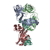
- Structure visualization
| Structure viewer | Molecule: MolmilJmol/JSmol |
|---|
- Downloads & links
-Download
| PDBx/mmCIF format | 4go6.cif.gz | 96 KB | Display | PDBx/mmCIF format |
|---|---|---|---|---|
| PDB format | pdb4go6.ent.gz | 73.5 KB | Display | PDB format |
| PDBx/mmJSON format | 4go6.json.gz | Tree view | PDBx/mmJSON format | |
| Others |  Other downloads Other downloads |
-Validation report
| Arichive directory | https://data.pdbj.org/pub/pdb/validation_reports/go/4go6ftp://data.pdbj.org/pub/pdb/validation_reports/go/4go6 | HTTPS FTP |
|---|
-Related structure data
| Similar structure data |
|---|


-Links
 PDBj
PDBj- Assembly
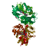
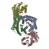
| Deposited unit | 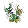
| ||||||||
|---|---|---|---|---|---|---|---|---|---|
| 1 | 
| ||||||||
| 2 |
| ||||||||
| 3 | 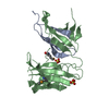
| ||||||||
| 4 | 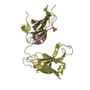
| ||||||||
| Unit cell |
| ||||||||
| Details | AUTHOR DETERMINED BIOLOGICAL UNIT: UNKNOWN. |
-Components
| #1: Protein/peptide | Mass: 4918.472 Da / Num. of mol.: 2 / Fragment: HCF-1 SAS1N, UNP residues 360-402 Source method: isolated from a genetically manipulated source Source: (gene. exp.) Homo sapiens (human) / Gene: Homo sapiens / Plasmid: pET21a, pET28a / Production host:  #2: Protein | Mass: 25228.830 Da / Num. of mol.: 2 / Fragment: HCF-1 SAS1C-NLS, UNP residues 1806-2035 Source method: isolated from a genetically manipulated source Source: (gene. exp.) Homo sapiens (human) / Gene: HCFC1, HCF1, HFC1 / Production host: #3: Chemical | ChemComp-SO4 /   Mass: 96.063 Da / Num. of mol.: 4 / Source method: obtained synthetically / Formula: SO4 Mass: 96.063 Da / Num. of mol.: 4 / Source method: obtained synthetically / Formula: SO4#4: Water | ChemComp-HOH / |  Mass: 18.015 Da / Num. of mol.: 33 / Source method: isolated from a natural source / Formula: H2O Mass: 18.015 Da / Num. of mol.: 33 / Source method: isolated from a natural source / Formula: H2OHas protein modification | Y | |
|---|
-Experimental details
-Experiment
| Experiment | Method: X-RAY DIFFRACTION / Number of used crystals: 1 |
|---|
- Sample preparation
| Crystal | Density Matthews: 3.14 Å3/Da / Density % sol: 60.83 % Description: THE STRUCTURE FACTOR FILE CONTAINS FRIEDEL PAIRS |
|---|---|
| Crystal grow | Temperature: 293.15 K / Method: vapor diffusion, hanging drop / pH: 8 Details: 2.23M ammonium sulfate, 5% Isopropanol, pH 8.0, VAPOR DIFFUSION, HANGING DROP, temperature 293.15K |
-Data collection
| Diffraction | Mean temperature: 110 K |
|---|---|
| Diffraction source | Source: SYNCHROTRON / Site: Photon Factory  / Beamline: AR-NW12A / Wavelength: 0.97892 Å / Beamline: AR-NW12A / Wavelength: 0.97892 Å |
| Detector | Type: ADSC QUANTUM 210 / Detector: CCD / Date: Jul 1, 2011 |
| Radiation | Protocol: SINGLE WAVELENGTH / Monochromatic (M) / Laue (L): M / Scattering type: x-ray |
| Radiation wavelength | Wavelength: 0.97892 Å / Relative weight: 1 |
| Reflection | Resolution: 2.7→50 Å / Num. obs: 42424 / % possible obs: 99.9 % / Redundancy: 7.3 % / Biso Wilson estimate: 59.5 Å2 / Rmerge(I) obs: 0.081 / Net I/σ(I): 31.3 |
| Reflection shell | Resolution: 2.7→2.75 Å / Redundancy: 7.3 % / Rmerge(I) obs: 0.637 / Mean I/σ(I) obs: 3.88 / % possible all: 100 |
- Processing
| Software |
| |||||||||||||||||||||||||
|---|---|---|---|---|---|---|---|---|---|---|---|---|---|---|---|---|---|---|---|---|---|---|---|---|---|---|
| Refinement | Method to determine structure: SAD / Resolution: 2.7→19.94 Å / Occupancy max: 1 / Occupancy min: 1 / σ(F): 0 / Stereochemistry target values: Engh & Huber / Details: THE FRIEDEL PAIRS WERE USED IN PHASING.
| |||||||||||||||||||||||||
| Solvent computation | Bsol: 36.6369 Å2 | |||||||||||||||||||||||||
| Displacement parameters | Biso max: 172.37 Å2 / Biso mean: 58.2096 Å2 / Biso min: 14.6 Å2
| |||||||||||||||||||||||||
| Refine analyze | Luzzati coordinate error obs: 0.37 Å / Luzzati d res low obs: 5 Å / Luzzati sigma a obs: 0.62 Å | |||||||||||||||||||||||||
| Refinement step | Cycle: LAST / Resolution: 2.7→19.94 Å
| |||||||||||||||||||||||||
| Refine LS restraints |
| |||||||||||||||||||||||||
| LS refinement shell | Resolution: 2.7→2.87 Å / Rfactor Rfree error: 0.025
| |||||||||||||||||||||||||
| Xplor file |
|
