Mass: 18.015 Da / Num. of mol.: 324 / Source method: isolated from a natural source / Formula: H2O
-
Details
Sequence details
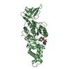
The protein used in this structure was directly purified from its source. The sequence of this ...The protein used in this structure was directly purified from its source. The sequence of this protein was determined based on the high resolution crystal structure electron density map. The electron density corresponding to the UNK region was not clear most likely due to structural disorder and thus the sequence could not be determined.
-
Experimental details
-
Experiment
Experiment
Method: X-RAY DIFFRACTION / Number of used crystals: 1
-
Sample preparation
Crystal
Density Matthews: 2.88 Å3/Da / Density % sol: 57.26 % / Description: Cuboid shape
Crystal grow
Temperature: 277.15 K / Method: vapor diffusion, hanging drop / pH: 7.5 / Details: 50mM HEPES, 12% PEG 3350
Resolution: 2.16→50 Å / Cor.coef. Fo:Fc: 0.957 / Cor.coef. Fo:Fc free: 0.934 / SU B: 4.505 / SU ML: 0.113 / Cross valid method: THROUGHOUT / ESU R: 0.174 / ESU R Free: 0.16 / Stereochemistry target values: MAXIMUM LIKELIHOOD / Details: HYDROGENS HAVE BEEN ADDED IN THE RIDING POSITIONS
Rfactor
Num. reflection
% reflection
Selection details
Rfree
0.21671
2983
5.2 %
RANDOM
Rwork
0.17515
-
-
-
obs
0.17735
54852
99.96 %
-
Solvent computation
Ion probe radii: 0.8 Å / Shrinkage radii: 0.8 Å / VDW probe radii: 1.2 Å / Solvent model: MASK
 Movie
Movie Controller


 Open data
 Basic information
 Components
 Keywords
 Function and homology information
 X-RAY DIFFRACTION /
  Authors
 India, 1items
  Citation
 Structure visualization
 Downloads & links
 Other downloads


 PDBj
PDBj

 Assembly
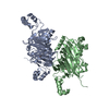


 Mass: 35.453 Da / Num. of mol.: 1 / Source method: obtained synthetically / Formula: Cl
 Mass: 22.990 Da / Num. of mol.: 1 / Source method: obtained synthetically / Formula: Na
 Mass: 138.121 Da / Num. of mol.: 2 / Source method: obtained synthetically / Formula: C7H6O3
 Mass: 63.546 Da / Num. of mol.: 2 / Source method: obtained synthetically / Formula: Cu
 Mass: 194.226 Da / Num. of mol.: 1 / Source method: obtained synthetically / Formula: C8H18O5 / Comment: precipitant*YM
 Sample preparation
 / Beamline: BM14 / Wavelength: 0.9787 Å
 Processing